1 Chapter 1
LAB 1
Animal Organization, Tissues and Integumentary System
Prepared by Dr. Jeff Ray, Dept. of Biology, UNA
OBJECTIVES
After completing these laboratory activities, you should understand/be able to:
- The hierarchical organization of the body and the four major tissue types in humans.
- The name, structure, basic function, and location in the body of the tissues examined in lab.
- Identify slides of the specific tissues prescribed by your lab instructor.
- List the properties of muscle tissues including striated/not, in/voluntary and branching/not.
- Label and identify the layers & components of the skin, the different burn classifications, and the skin cancer model.
Introduction
The basic building blocks of living organisms are cells, with an estimated 15-50 trillion cells in the human adult body. Groups of specialized cells along with their extracellular matrix that carry out a specific function are tissues. Two or more tissue types which collectively function as a unit are organs, and organs function cooperatively as organ systems. The 11 organ systems in humans contribute to maintaining homeostasis of the individual organism. Thus, the hierarchical organization of the body is cells→ tissues→ organs→ organ systems→ organism.
Among the more than 200 types of cells in humans, many cells (and thus tissues) are often visually distinguishable by their overall shape, the presence/absence of a nucleus, the amount of fibers in the cell, the amount of extracellular material or spaces between cells, and the presence/absence of cilia or other cell projections. The shapes of cells are invariably linked to their function (i.e. form and function are correlated). Although most cells have a nucleus, exceptions include red blood cells, which lack a nucleus (anuclear), while muscle cells are multi-nucleated. Often, the nucleus takes a similar shape to the overall cell shape. There are dozens of specific tissues in humans, within four major types (classes): epithelial, connective, muscle, and nervous. Different combinations of these tissues make up our organs and organ systems.
Histology is the study of microscopic anatomy of cells and tissues of organisms. Traditionally, a light microscope is used to observe cells and tissues at <1000X magnification. In order to be visualized, the specimen must be chemically preserved, embedded (dehydrated & infused with wax) sectioned (into thin slices), and stained onto a slide (fixed-embedded-sectioned-stained). Normally, the nucleus, fibers, cell margins, cilia, villi, intercalated disks and other components are contrasted by the staining process.
* List the levels of organization: _________→ _________→ ___________→ __________ organism
* What are the 4 basic types of tissues found in animals? __________ ,__________, _______,________
Basic Instructions: Microscope Usage
- Correctly place slide on the microscope stage
- Using knobs, center & focus image (1st coarse, then fine focus) using LOW power objective lens first
- Zoom to 10x or 40x to view at ideal magnification (use lab manual images for comparison)
- Only use fine focus after zooming from low power
- Do not use the oil immersion setting for the (highest power) objective lens
– For each tissue: the specific name & its location in the body is listed inside the lid of the slide box
– Many other tissues are present in most slides, which must be distinguished from the tissue of interest
– Ask the instructor to verify your tissue identification, compare to book images or look up images
Epithelial Tissue
Epithelial tissue covers the body, lines its cavities, and forms glands. It contains one or more layers of closely adhering cells that cover external and internal surfaces like the skin, lungs, and intestines. One surface is exposed (apical: faces the environment), while the other surface is normally anchored to a basement membrane (basal surface). Epithelial tissues mainly function in either protection, secretion or absorption. They lack blood vessels (avascular); cells receive oxygen and nutrients from blood vessels that are adjacent to them.
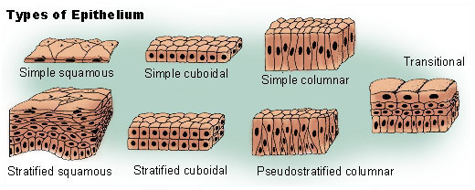
The multi-part name of epithelial tissues generally refers to the number of layers present and the shape of the cells. Layers may be single (simple) or ≥ (stratified). Three common shapes are squamous, cuboidal, and columnar. The combinations of these names describe most epithelial tissues: simple columnar, stratified squamous, etc. Pseudostratified tissues appear to be layered, but all cells reach the basement membrane, and are technically one layer thick. Cellular extensions including cilia and microvilli are present some in epithelial tissues.
*Where are epithelial tissues specifically found in the body? ___________________________________
*Epithelial tissues are named based on what two major characteristics? _____________, ____________
*What do these terms mean in reference to epithelial tissue?
Simple – Stratified – Pseudostratified –
Squamous – Cuboidal – Columnar –
1.)Simple Squamous Epithelium
Function: Absorption (rapid diffusion)
Locations: Linings of lungs and blood vessels
Tissue to examine: 1.) Human Lung
Simple squamous epithelium lines spaces in the body which primarily function in absorption (rapid diffusion). The single layer of scale-like cells facilitate easy movement across the surface area. Examine a tissue slide from the lining of the human lung. The lining of blood vessels, the heart, and portions of the respiratory, urinary and reproductive tract also contain this tissue.
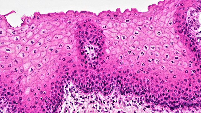
2.)Stratified Squamous Epithelium
Function: Protection
Locations: Epidermis, mouth, throat
Tissue to examine: 2.) Human Skin
Stratified squamous epithelium contains multiple cell layers which primarily function in protection from abrasion. The layers near the surface (superficial) are flattened and older, while deeper layers have a cuboidal or columnar shape and are more recently formed. You will examine a tissue slide from the human epidermis; try to count the approximately 30 cell layers. The outermost tissues of the skin (epidermis), mouth, esophagus, anal canal, and vagina contain this tissue.
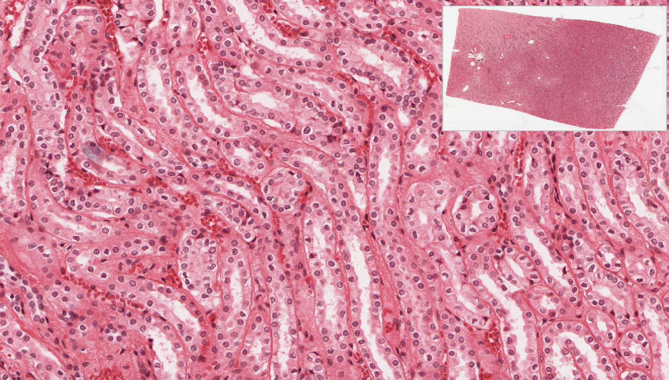
3.)Simple Cuboidal Epithelium
Function: Absorption of molecules
Locations: Kidneys, ducts of glands
Tissue to examine: 3.) Kidneys
Simple cuboidal epithelium contains a single layer of cube-shaped cells anchored to a basement membrane and primarily functions in absorption. The outer surface often contains microvilli (not visible with light microscope) that increases the surface area for absorption. Examine the tissue slide from the human kidney; notice the centrally-located nucleus that is roughly the same shape as the cell overall. The tubules of the kidneys, liver, and the ducts of various glands contain this tissue.
4.)Simple Columnar Epithelium
Function: Absorption of nutrients
Locations: Stomach, intestines (digestive tract)
Tissue to examine: 4.) Stomach (pyloric region)
Appears as a row of tall and narrow cells each with a single nucleus near the base; nucleus takes the overall shape of the cell. Primarily functions in nutrient absorption. Wavy folds increase the surface area for absorption. Examine human stomach tissue; identify goblet cells which secrete mucus (may stain light blue). Mucus lubricates surface and protects from stomach acid & enzymes. The digestive tract is lined by this tissue.
What is the function of the mucus?
______________________________________
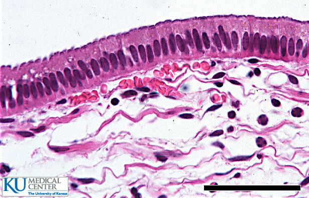
5.) Pseudostratified Ciliated Columnar Epithelium
Function: Sweeps impurities from airway, protects
Locations: Trachea (windpipe), bronchi
Tissue to examine: 5.) Trachea
Pseudostratified ciliated columnar epithelium looks layered due to cell nuclei at different levels, but each cell reaches the basement membrane (pseudo = false). Hair-like projections called cilia act as tiny brooms to sweep debris trapped in mucus up and out of the airway. This tissue primarily functions in clearing the airway of dust, bacteria, and other debris that could otherwise enter the lungs. Smoking coats cilia in many fine particles. The wavy folds of the tissues increase the surface area for absorption. Examine the tissue slide from the trachea; identify the goblet cells which secrete mucus (may be stained light blue). Try to identify at least five other tissues found in slide 5.
*Where is this type of tissue found in vertebrates?
______________________________________
*What is the function of the cilia?
______________________________________
*How might the cilia be affected by smoking?
______________________________________
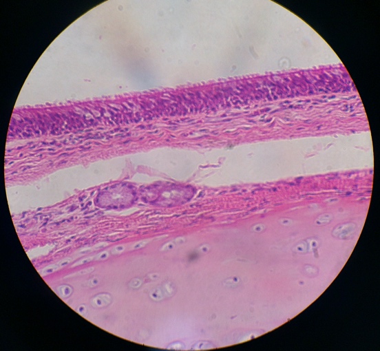
Table 1: EPITHELIAL TISSUE
|
Type |
Structure |
Function |
Location |
|---|---|---|---|
| Simple Squamous | rapid diffusion | ||
|
Stratified Squamous |
outer layers flat, inner layers cuboidal | ||
| Simple Cuboidal | secretion & absorption | ||
| Simple Columnar | Taller than wide, nucleus at base | digestive tract, uterus | |
|
Pseudostratified ciliated columnar |
protection & secretion, sweeps substances |
*Complete the missing information in the table above.*
Connective Tissue
Connective tissue is the most variable and widespread tissue in the body. Specific types vary in their amounts and ratios of cells, fibers, and extracellular matrix. Cells may be numerous or few, fibers vary in amount and type (elastin or collagen), and the extracellular matrix may be limited or extensive and contain gelatinous (cartilage) or rigid materials (as in bone). Connective tissues function in support, binding, insulation, protection, and friction reduction, among other purposes. Note: fibers in connective tissue are non-cellular fibers made of proteins; fibers in muscle tissue are whole cells- do not confuse connective tissue “fibers” with the fibers of muscle tissue.
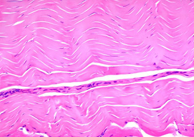
6.)Dense regular connective tissue
Function: Connect bones and muscles
Locations: Tendons, ligaments
Tissue to examine: 6.) Tendon
Dense regular (fibrous) connective tissue is tightly filled with wavy parallel collagen fibers, and the tissue has an appearance somewhat like woven rope. The nuclei are dark-stained and appear pill-shaped, with no extracellular spaces visible within the tissue. Dense fibrous tissue imparts strength to tendons and ligaments, primarily in a single direction; forces applied parallel to the tissue fibers more easily tear the tendon or ligament.
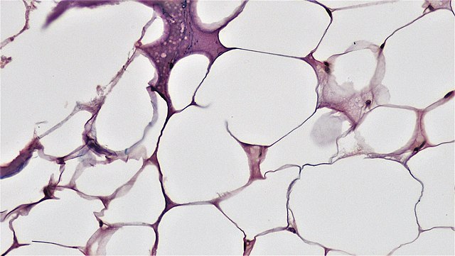
7.)Adipose tissue
Function: Insulation, protection, and energy storage
Locations: Beneath skin, surrounds organs, breasts, hips, gut
Tissue to examine: 7.) Adipose tissue
Cells of adipose tissue are filled with fat and triglycerides, which do not stain well, thus the cells appear mostly empty. The nuclei are pushed to the side of the cell and the cell edges have a dark margin. Nuclei are visibly stained along the cell margins and there is no extracellular spaces visible within the tissue. Adipose tissue functions in storing energy, insulation, and cushioning around organs and other structures.
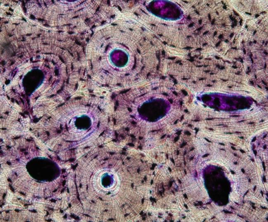
8.)Compact bone
Function: Support and protection, mineral storage
Locations: Skeleton
Tissue to examine: 8.) Compact bone
Bone tissue is one of the most distinct tissues, and is obviously found in the skeleton. Compact and spongy bone are the two main types; both varieties are found in most bones (e.g. femur); compact bone makes up the majority of the skeleton. A system of interconnecting vascular canals (haversian systems) contain the blood supply for living cells (osteocytes), which are embedded within a calcified matrix of extracellular materials. The functional unit of compact bone is an osteon, which appear as adjacent tree rings. Examine the slide and the model of bone tissue. Be able to find the osteon, central canal, and osteocyte.
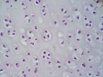
9.)Hyaline Cartilage
Function: Support and protection, minimizes friction
Locations: Nose, trachea and bronchi, ends of bones, ribcage, intervertebral disks
Tissue to examine: 9.) Hyaline cartilage
Hyaline cartilage is a connective tissue with an extracellular gel-like matrix containing few/no visible fibers, scattered cells called chondrocytes, and overall appear less organized than bone. This tissue supports, protects, and minimizes friction where bones meet. Hyaline cartilage is found in the nose, trachea, ribcage, intervertebral disks and covers the ends of bones. The two other cartilage types (elastic, fibrous) are not examined in this lab.
10.)Blood
Function: Transport of nutrients and wastes
Locations: In blood vessels and heart
Tissue to examine: 10.) Blood smear
Blood is a connective tissue which contains a mixture of liquids, cells, and cellular fragments. The liquid remains unstained, while the vast majority of cells are red blood cells, which appear as donut-like disks- no nuclei are present. White blood cells (WBCs) are far fewer in number, but noticeably larger and distinctly stained. The five main types of WBCs are identified based on the shape of their nuclei. WBCs function in fighting infections. Platelets are cell fragments that are essential to blood clotting.
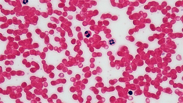
Blood smear dominated by RBCs, zoom to see WBCs & platelets
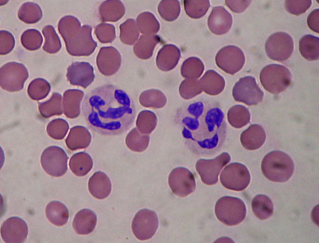
Blood tissue showing neutrophils (WBCs).
Table 2: CONNECTIVE TISSUE
|
Type |
Structure |
Function |
Location |
|---|---|---|---|
|
Dense fibrous |
|
||
|
Adipose |
Beneath skin, surrounds organs, hips, breasts, stomach | ||
|
Compact bone |
Support & Protect | ||
|
Hyaline Cartilage |
Scattered cells, Gel-like matrix |
Nose, trachea & bronchi, bone ends, ribcage | |
|
Blood |
Plasma, RBCs, WBCs, platelets |
*Complete the missing information in the table above.*
Muscle Tissue
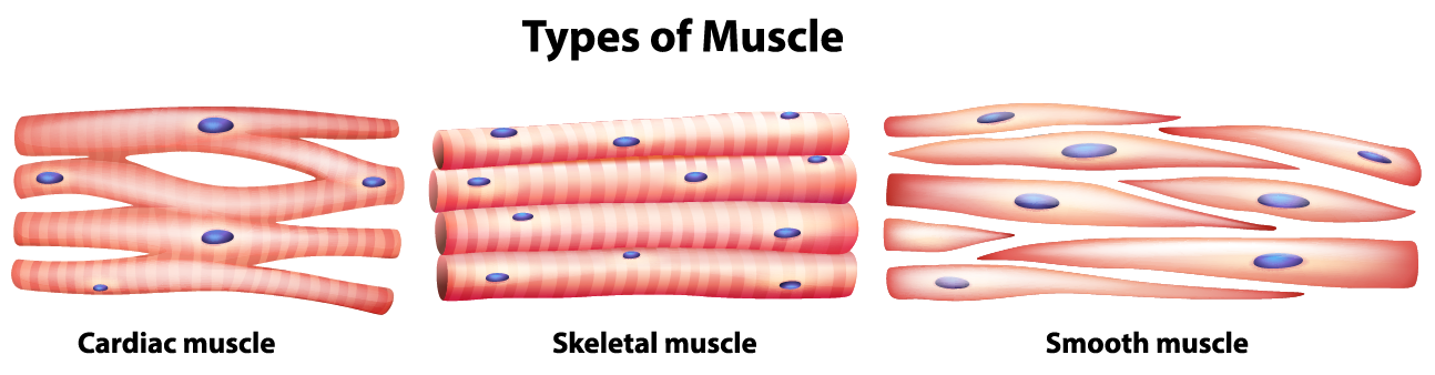
Muscle cells are long & thin and called myocytes, which are filled with fibers that contract by shortening. When the tissues contract, they function in moving the body or moving substances like blood or food through the body. Skeletal, cardiac, and smooth muscle tissues vary in shape from one another; the structure gives insight as to how the muscle functions when contracting. Muscle tissue also varies in being not/striated and in/voluntary; one or more nuclei are present.
*All muscle tissues are composed of cells called ______________________________________.
*What are the three types of muscle tissues? ________________________________________________
11.)Skeletal muscle tissue
Function: Movement (voluntary)
Locations: Skeletal muscles like biceps, deltoids
Tissue to examine: 11.) Skeletal muscle
Skeletal muscle tissue contains long, tube-shaped cells filled with parallel fibers containing actin and myosin protein fibers. These fibers alternatively do/not overlap, causing the tissue to have a striated (striped) appearance like a candy cane. The nuclei are numerous (multi-nucleated), darker, and pushed to the margins of the cells. There are no extracellular spaces visible within this tissue. There are more than 600 skeletal muscles that function in moving the body; these are under voluntary (conscious) control of the nervous system. Obtain a slide of skeletal muscle and be able to identify the fibers, nuclei, and striations. Adjust the contrast setting to better visualize the striations.
*Is skeletal muscle voluntary or involuntary? _________________________________
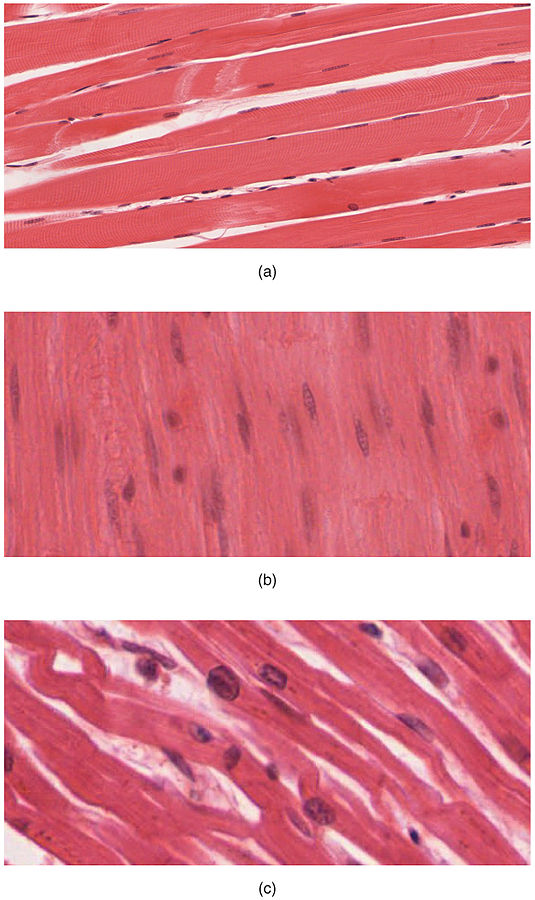
Top to bottom: skeletal, smooth, cardiac
12.) Cardiac muscle tissue
Function: Pumps blood
Locations: Heart
Tissue to examine: 12.) Cardiac muscle tissue
Cardiac muscle is restricted to the heart and has an appearance unique among muscle tissue- it is striated and branching with one nucleus per cell. The branching cells are interconnected so the heart may function as a unit. The cell junctions are held together by zipper-like structures called intercalated disks, which allow substances and electrical current to pass seamlessly among cells. There are no extracellular spaces visible within the tissue. Cardiac muscle is involuntary (unconscious).
13.) Smooth muscle tissue
Function: Move substances through the body organs
Locations: Viscera (digestive, endocrine and reproductive organs) in body cavities
Tissue to examine: 13.) Smooth muscle tissue
Smooth muscle makes up the wall of internal organs like the stomach; it is restricted to the viscera and blood vessels. It has a different appearance from other muscle tissues- it is not striated, is not branched and contains one central nucleus per cell, which normally stains darker than fibers. The cells are filled with fibers, appearing narrow at the ends and wider in the middle giving a spindle-shaped appearance. There are no extracellular spaces visible within the tissue. Smooth muscle is under involuntary (unconscious) control. Smooth muscle is found in the walls of the trachea, esophagus, stomach, intestines, blood vessels, and urinary bladder.
Table 3: MUSCLE TISSUE
|
Type |
Striated (Yes/No) |
In/Voluntary |
Branching (Yes/No) |
|---|---|---|---|
| Skeletal | |||
|
Smooth |
|||
| Cardiac |
* Complete the missing information in the table above.*
*When finished examining the 13 tissue slides, organize them in the slide box in the correct order. If any slides are damaged, please notify your instructor. Turn off and unplug your microscope, put the objective lens on low power setting, and place the plastic cover on the microscope.*
Nervous Tissue
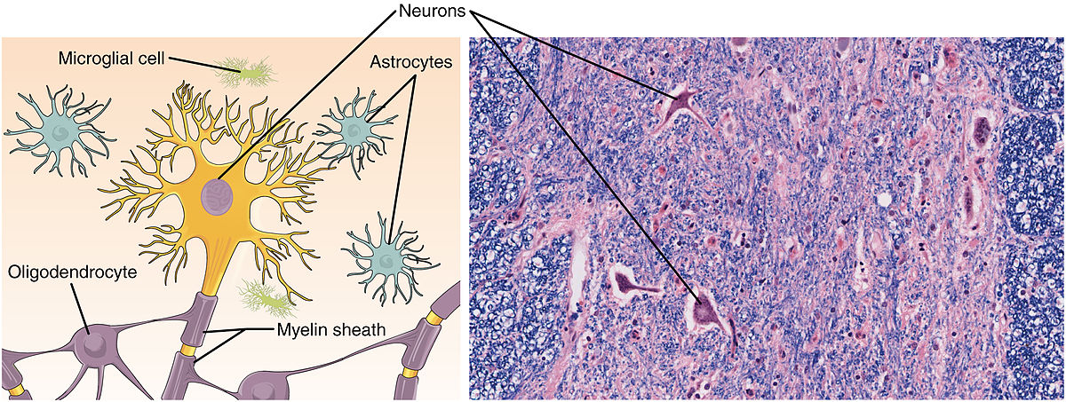
No nervous tissue slides will be examined in lab, but several types of nervous tissues are found in the brain, spinal cord, and nerves. Nerve cells that carry electrical impulses are neurons, while the numerous supporting cells are neuroglia; several sub-ypes of neurons and neuroglia are recognized.
*Where is nervous tissue found in vertebrates? ____________________________________________
*What are the two main types of cells in nervous tissue? _____________________________________
Integumentary System
The integumentary system includes the skin, hair, and nails which have multiple functions in maintaining homeostasis, including protection and temperature regulation. The skin is composed of the epidermis and the dermis. The tissue layer underlying these two layers is the hypodermis (=subcutaneous layer) which has a different origin during embryo development and therefore, is not considered a “true” layer of the skin. From superficial (outer) to deep (inner), the layers are epidermis—dermis—hypodermis. The epidermis contains stratified squamous epithelial tissue that is constantly being replaced. There are variably shaped layers of cells, called stratum (layers a-e), the outermost of which are dead and contain a waterproof coating. The dermis is thicker and primarily connective tissue, but also contains glands, nerves, and blood vessels (epidermis is avascular). The hypodermis (layer III) is adipose tissue and the layer into which certain medical injections are given (e.g. insulin shots).
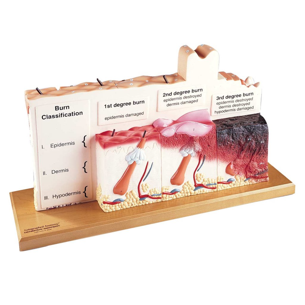
Examine the human skin model, including burn pathologies on the back of the model. Note the following:
– 3 skin regions: palm, armpit, scalp – 3 distinct layers (I-III): epidermis, dermis, hypodermis
– Strata (a-e) of the epidermis – Blood vessels, nerves, hair root, etc. in dermis
– Adipose tissue (g) of hypodermis – Depth, damage of 1st, 2nd and 3rd degree burns
* Which region of skin is the thickest: palm, armpit or scalp? _______________________
*Name the main tissue types:
Epidermis: __________________ Dermis: _________________ Hypodermis: ______________
* Which layer is thickest: epidermis, dermis or hypodermis? _____________________
*Which degree of burn destroys the epidermis and damages the dermis (often the most painful)? ______
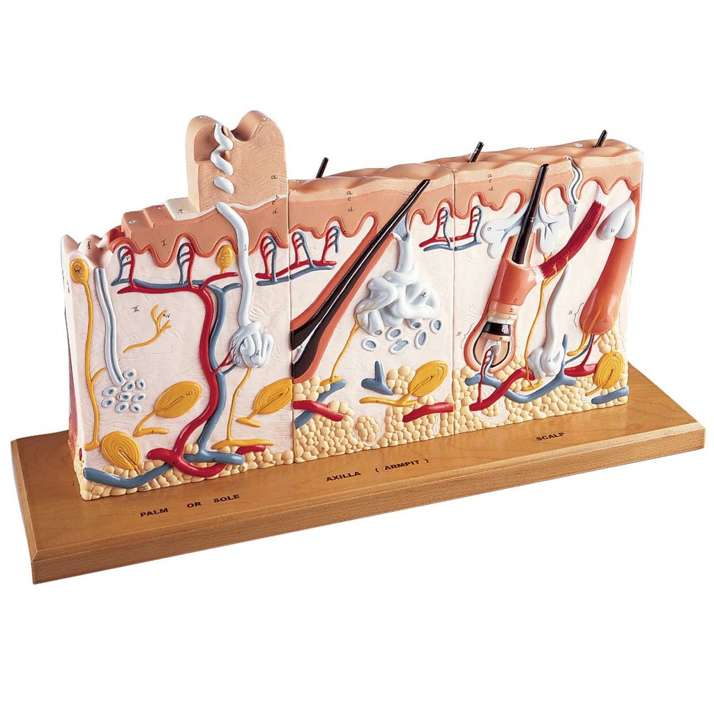
Lab model of the skin. Note I: Epidermis, II: Dermis, III: Hypodermis. Nerve receptors in various locations of the dermis detect light touch, deep pressure, pain, hot, and cold.
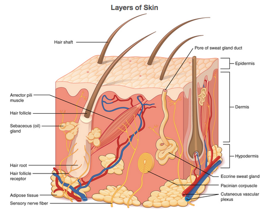
Labelled model of the skin.
Skin cancer is the most common type of cancer, due to the (1) constant exposure to the environment, (2) frequent cell division, and (3) large size of this organ system. Of the three main types, malignant melanoma is the rarest, but the most deadly (other two: squamous cell carcinoma, basal cell carcinoma). The six zones on the model show the progression of untreated melanoma: cells multiply, deepen into the skin, and metastasize (spread elsewhere in the body). The American Cancer Society guide for early detection of melanoma via self-exam uses the “ABCDE rule” to assist individuals in monthly self-exams of skin discoloration that might be malignant, to aid in early detection by a dermatologist. A= asymmetry (unequal halves), B = border irregular, C = color variable (differing shades), D = diameter >1/4”, E = evolving (changing).
Examine the skin cancer model, note the following:
– 6 zones, read left to right, front to back
– Progressive deepening of tumor into the skin
– Metastasis into the blood vessels/lymph
– Variable shape of tumors on base of model
*Name the deadliest skin cancer (on model):_____________________________
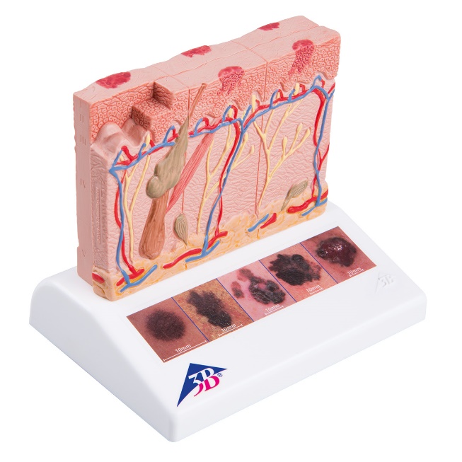
 J15 Skin Cancer Model
© 3B Scientific GmbH, Germany, 2019, www.3bscientific.com
Image Credits:
Simple Squamous By https://en.wikipedia.org/wiki/Simple_squamous_epithelium#/media/File:Illu_epithelium.jpg
Stratified squamous By https://commons.wikimedia.org/wiki/File:Epithelial_Tissues_Stratified_Squamous_Epithelium_(40230842160).jpg
Simple cuboidal By https://histology.medicine.umich.edu/sites/default/files/images/slides/1epithelial.jpg
Simple columnar lecannabiculteur.free.fr/SITES/UNIV%20KANSAS/instruction/medicine/anatomy/histoweb/gitract/large/Gi39.jpg
Pseudostratified ciliated columnar By Assassin3577 – Own work, CC BY-SA 3.0, https://commons.wikimedia.org/w/index.php?curid=26594055
Dense Regular Connective By http://medcell.med.yale.edu/histology/connective_tissue_lab.php
Adipose https://commons.wikimedia.org/wiki/File:Connective_Tissue_Adipose_(41066514324).jpg
Bone By https://commons.wikimedia.org/wiki/File:Compact_bone_-_ground_cross_section.jpg
Cartilage By https://commons.wikimedia.org/wiki/File:Cartilage01.JPG
Blood https://commons.wikimedia.org/wiki/File:Connective_Tissue_Human_Blood_Leukocyte_Survey_(26921278957).jpg
Neutrophils By https://commons.wikimedia.org/wiki/File:Segmented_neutrophils.jpg
Muscle tissues By OpenStax College – Anatomy & Physiology, Connexions Web site. http://cnx.org/content/col11496/1.6/, Jun 19, 2013., CC By 4.0, https://commons.wikimedia.org/w/index.php?curid=30015032
Muscle tissues By http://www.scientistcindy.com/uploads/8/5/1/2/85124478/muscle-tissue1_orig.png
Nervous tissue By By OpenStax College – Anatomy & Physiology, Connexions Web site. http://cnx.org/content/col11496/1.6/, Jun 19, 2013., CC BY 3.0, https://commons.wikimedia.org/w/index.php?curid=30131294
Skin model front and back https://denoyer.com/products/human-skin-series
Skin (labeled) By OpenStax College – Anatomy & Physiology, Connexions Web site https://cnx.org/contents/FPtK1zmh@8.108:RxywCGkA@6/Layers-of-the-Skin
Melanoma model used with Permission of 3B Scientific, images from https://www.a3bs.com/skin-cancer-model-1000293-j15-3b-scientific,p_54_316.html
BI 102 Lab Worksheet: Tissues Name _________________________________ Section _______
1. What are the 4 major types of tissues found in animals?
2. Name 1 specific location in the body you would find the following epithelial tissues:
a. simple squamous epithelial _________________ b. stratified squamous epi.__________________
3. Find pseudostratified ciliated columnar epithelial tissue (slide #5) with your microscope. Put your pointer on this tissue and have your instructor verify it by initialing here ______________
4a. Slide #5 Sketch AND LABEL this tissue:
4b. List the other tissues present on slide #5:
______________________, ___________________
______________________, ___________________, ______________________
5. Name 3 specific types of connective tissues (see Table 2)
______________________, _______________________,_______________________
6. Sketch the following tissue: Compact Bone (slide #8)
7. Find a White Blood Cell (slide #10 and sketch it (ZOOM in to 40X- include nucleus shape)):
8. What are the 3 basic types of muscle tissue? (slides #11-13)
9. Which muscle tissue is striated, branching and not under conscious control (involuntary; Table 3)?
10. Sketch AND Label the two “true” layers and the third underlying layer of the skin→

.jpg)
.jpg)