7 Chapter 7
LAB 7
The Senses
Prepared by Jason R. Jones & Dr. Jeff Ray, University of North Alabama
Introduction
To detect our environment, the body uses receptors to detect various stimuli in the internal and external environment to help maintain homeostasis. These stimuli are transmitted as electrical impulses through nerves to the brain for processing/interpretation. Receptors include photoreceptors (visible light), chemoreceptors (detect various molecules like CO2), thermoreceptors (hot or cold), mechanoreceptors (including pressure & in the ear for hearing and balance), and pain receptors.
In today’s lab, you will do activities that measure the five traditional senses for vision, smell, taste, sound, touch, and also temperature.
* What type of receptors detect light? _________________smell & taste? ___________________
sound? _________________temperature? ____________________
Eyes and Vision
Eyes are special sense organs for detecting wavelengths of visible light using photoreceptors. Humans have a camera-type eye that focuses light rays on the retina to form an image detected by the brain. The cornea is the outer surface known as the ‘window of the eye’. The pressure in the front portion of the eye is maintained by the water-like aqueous humor. The iris is the colored portion of the eye, while the pupil can constrict or dilate to control the amount of light passing through it into the eye. A single lens focuses the image onto the retina. Before reaching the retina, light passes through the jelly-like vitreous humor. Light rays strike the retina, which contains two main types of photoreceptors, rods and cones. Rods are more numerous and detect the presence/intensity of light. The cones are fewer in number and each of three types detect different wavelengths (colors) of light; the combinations of these types forms the various colors we perceive.
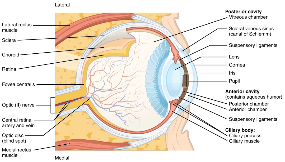
*What is the path of light from outside the eye to where it is detected by photoreceptors?
(word bank: aqueous humor, cornea, lens, pupil, retina, vitreous humor)
____________→ ___________→ ____________→ ____________→ ___________→ ___________
Check the boxes when you complete each exercise. Do the visual acuity and astigmatism together using the wall charts.
Visual Acuity
The Snellen chart for visual acuity presents a limited number of letters in lines of decreasing size. The line with letters that are marked 20/20 represent the smallest letters that a person with normal acuity should be able to read with 100% accuracy from 20 feet. The different sizes of letters in the other lines represent rough approximations of what a person of normal acuity can read at different distances. For example, the line that represents 20/200 vision would have larger letters so that they are legible to the person with normal acuity at 200 feet. The 20/200 value (or worse) is considered “legally blind” if not corrected with lenses.
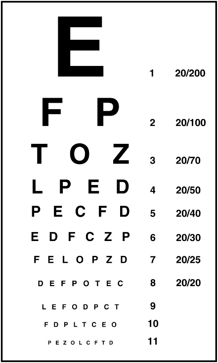
____ To Test For Visual Acuity:
Use the Snellen eye charts^ on the wall, stand 20’ away. Do not squint. If you wear glasses, test both with
and without your glasses. Do not remove contacts to test.
1. Test 1 eye at a time, do the left eye first. Have your partner stand at the eye chart and point to each line.
2. Close or cover your right eye. Read each line from the top to the bottom of the chart.
3. The bottom-most line you read with 100% accuracy gives an approximation of your visual acuity versus what is considered normal vision (20/20).
4. Repeat for your right eye, close or cover your left eye.
5. Record your results below, use the second set of blanks after removing your glasses, if you wear any.
a. Left eye 20/_____b. Right eye 20/_____c. Left eye 20/____d. Right eye 20/____
^ This test is very primitive as compared to the advanced instruments an optician can use to determine your visual acuity and correct deficiencies in vision.
Explanation: If your vision is 20/40, you can see at 20’ what can be seen from 40’ in a person with normal vison; you have worse than average vision. If your vision is 20/15, you can see at 20’ what a person with normal vision can only see clearly from 15’ (closer up), you have better than average vision.
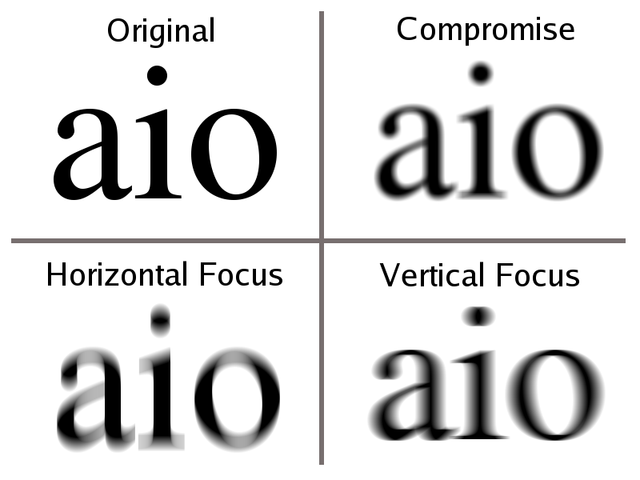
Astigmatism
Astigmatism is a refractive error in the eye caused by an unevenness in the cornea or lens that can blur or distort vision at all distances. These surface abnormalities tend to bend light irregularly based upon the convex and/or concave shape of the eye surface. The result is the scattering of light and a lack of clarity in a portion of the field of vision. Astigmatism is normally corrected by glasses, contacts or surgery. If left uncorrected, astigmatism can lead to headaches, fatigue, squinting and pain in the muscles around your eye.
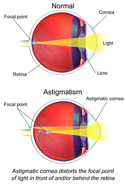
____ To Test for Astigmatism: Use the astigmatism wheel charts^ on the wall, stand 20’ away. Do not squint.
1. Test 1 eye at a time, do the left eye first.
2. Close or cover your right eye. Focus your vision on the center circle, while examining each line that radiates off of it. If you do not have astigmatism, the lines will appear sharply focused and equally dark. You may have astigmatism if some lines appear sharp and dark, while others are blurred and lighter.
3. Test the right eye following the same steps. Record your observations below.
Astigmatism a. Left eye: Yes / No b. Right eye: Yes / No
^ This test is very primitive as compared to the advanced instruments an optician can use to diagnose astigmatism.
*Which part(s) of the eye are abnormal in an astigmatism?
________________________________________ ______________________________________
Superimposition
Superimposition refers to the process by which the separate images detected by each eye are combined into a single three-dimensional image by the brain. Since each eye has a separate field of view, the two images differ slightly, but your brain combines them into one cohesive image. For this activity, you are going to trick your brain by having your eyes send conflicting information about what you are seeing.
____ To Demonstrate Superimposition: Use the hollow paper tube at your table.
1. Find an object on the wall, such as an exit sign, as your object to view.
2. Close your left eye and keep your right eye open. Raise the tube and hold it up against your right eye and view the wall object through the tube. Your field of view should be the sign through the tube.
3. Raise your left hand 12” in front of your left eye alongside the tube with your palm facing away, while holding the tube steady.
4. Close your right eye and open your left eye. You field of view should be the back of your hand.
5. While leaving the tube and your hand in place, open both eyes and note what you see. Slide your hand up and down the tube with your palm facing away to highlight this optical illusion.
6. Switch sides with the tube if the effect is minimal- one eye is often dominant and the brain prefers info from that side.
*Describe what you see when looking through the tube with both eyes open:
______________________________________________________________________________
Explanation: one is generally unaware of superimposition under normal conditions- the brain combines the separate images into a single visual perception which generally does not conflict. This exercise created a situation called a binocular rivalry in which one is directly aware of the two separate fields of view.
Afterimages
We see because the rods and cones are generating nerve impulses that are transmitted to the brain. Occasionally, the brain retains an image even after the impulses have stopped (afterimages). Positive afterimages are when the bright parts of the object remain bright & the dark parts remain dark. Negative afterimages are when the bright parts of the object appear dark & the dark parts appear bright.
____To Demonstrate Afterimages: Use fluorescent strips, black paper, white paper.
1. Center the fluorescent green strip of paper on the black construction paper and stare intently at the green strip for 30 seconds without shifting the eyes. Then have your partner quickly slide a white sheet of paper over the green strip. Record what afterimage you see, if any (possibly a flash of different color).
* Green afterimage- describe what you see:______________________________________________
Repeat the procedure using the fluorescent orange strip of paper.
* Orange afterimage- describe what you see:______________________________________________
The Blind Spot of the Eye
The blind spot occurs where the optic nerve exits the retina. Since the surface of the optic nerve lacks any photoreceptors, when light rays strike this portion of the retina, no image is detected due to the absence of both rods and cones.
____ To Demonstrate the Blind Spot: Use the strip of paper which has a small circle and a cross.
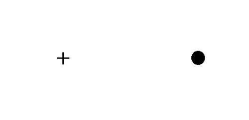
1. Test 1 eye at a time, do the left eye first.
2. Hold the paper strip at arm’s length with the cross directly in front of your left eye and the circle to the left. Close your right eye.
3. Stare only at the cross, do not let your left eye wander from it (the circle will be in the periphery).
4. Slowly move the paper toward your eye until the circle disappears.
5. Repeat as needed to find the blind spot (adjust it closer/further to find the exact distance it disappears).
6. With your partner’s help, measure the distance from your eye to the paper using a meter stick, record in cm.
7. Repeat procedure with your other eye, record distance.
*Blind spot distance: Left: ___________ cm Right: ___________ cm
*Which part of the eye which lacks rods & cones and causes the blind spot? ______________________
Explanation: one is generally unaware of the blind spot under normal conditions- the brain interprets what we perceive in part on past experience and essentially, fills in missing gaps. This exercise created an artificial situation in which one is directly aware of how perception if constrained by eye anatomy.
Accommodation of the Eye
When the eye accommodates to see objects at different distances, the shape of the lens changes. When you are looking at a distant object, the lens is flatter. When you are looking at a closer object, the lens becomes more curved (rounded). The lens shape is controlled by the ciliary muscles attached to it. The rounded lens bends light more to accommodate the closer image to fit into the eye. The ciliary muscles and the elasticity of the lens determines how well the eye can accommodate, and lens elasticity decreases with age, a condition called presbyopia. Presbyopia is the reason many older people need reading glasses to see near objects. The near point is the closest distance at which your eye can change shape to bring an object into focus (accommodation).
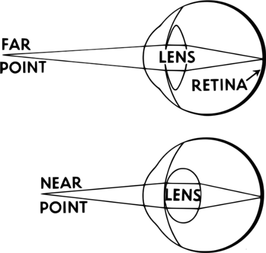
Figure: Light from a distant object and light from a near object brought to a focus on the retina. Notice how the lens is flatter for distant objects and is rounder for close objects. The lens rounds to accommodate near objects.
____ To Demonstrate Accommodation/ Determine Near Point:Use a pencil and meter stick.
1. Test 1 eye at a time, do the left eye first.
2. Hold the pencil at full arm’s length with the pencil tip pointing towards the ceiling. Close your right eye.
3. Focus on the pencil tip, and slowly move it toward your left eye until the end is out of focus.
4. Repeat as needed to find the distance (adjust closer/further to find the exact distance it goes out of focus).
5. With your partner’s help, measure the distance from your eye to the pencil using a meter stick, record in cm.
6. Repeat procedure with your other eye, record distance.
7. Compare your near point accommodation with values from the chart below. Determine the “age” of your eye.
*Near point accommodation: Left: ___________ cm Right: ___________ cm
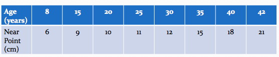
*What is the “age” of each eye? Left: ___________ Right: ___________
*Which part of the eye changes shape to accommodate? ______________________________________
Color Blindness
Color blindness is the decreased ability to see color or color differences and is due to a lack of functional cones (photoreceptor proteins) on the retina. Although a given color wavelength of visible light strikes the retina, it is not detected since there are no functional cones to detect it, no stimulus is sent to the brain and the person is ‘color blind.’ The genetic condition is more common in males versus females since the genes responsible for most forms of color blindness are on the X chromosome (females have two copies, so if one is defective, and the second copy functions, the individual would not be color blind). Diagnosis is typically with the Ishihara color test; but a number of other testing methods have been developed.
__To Test for Color Blindness: Use the Ishihara’s Tests for Colour Deficiency book at the table.
1. Observe plates 1-14 with both eyes. Plates will contain numbers, lines or patterns distinct from the surrounding dots. Count the number of plates you can clearly read.
• Plate No. 1 is a control and should be readable as ‘12’ by all persons with basic visual abilities.
• Plates 1-11 determine normal/defective color vision. Plates 12-14 determine the type and degree of color vision deficiencies.
• Analyze your results and look up explanations in the small booklet provided with the Ishihara book.
* Your Color vision score (#plates read): _________________________
10 or more plates: normal color vision7 or fewer: color vision deficient. See booklet for explanations
* Is color-blindness caused by a lack of functional rods or cones in the retina? _________________
Sense of Touch and Sense of Hot & Cold
The sensory receptors in skin respond to touch, pain, temperature, and pressure. Each of these stimuli have different receptors that detect them (hot & cold receptors are also separate), as well as free nerve endings, which respond to pressure, pain, and temperature. These messages are sent as electrical stimuli to the brain and processed.
____ To Measure Sense of Touch:
Use the grey calipers to pressure the skin and measure the distance between points. You will be testing the subject’s ability to discriminate between the two points of the calipers at the four different locations listed below.
*A laboratory partner is required for this procedure. Enter your data, not your partner’s data.
1. The subject must be seated with eyes closed. Hold the points of the calipers 20 mm apart on the given skin area, with both points gently touching the subject. Ask the subject whether the experience involves ‘one or two?’ touch sensations.
2. Move the calipers a few mm closer each time until the subject can only discriminate a single point. Record the shortest distance between the caliper points the subject can make a two-point discrimination.
3. Record the data in the blanks below. Remember, the subject cannot look at the calipers when being tested.
a. Forearm: ____________ mm
b. Back of the neck: ____________ mm
c. Index finger: ____________ mm
d. Back of the hand: ____________ mm
*Which area had the smallest two-point discrimination? ______________________________________
This area would have the most nerve endings allowing for the greatest sense of touch.
____ To Measure Sense of Hot & Cold:
Use the three large beakers containing hot, cold & room temperature tap water (on cart).
1. Immerse your left hand in the ice water beaker and your right hand in the hot water beaker for 30 seconds.
2. Simultaneously move both hands very quickly to the room temperature water in the middle beaker.
3. Record the immediate sensation you feel in both your L and R hands. Switch the beakers if you have time.
Temperature perceived in: a. Left hand: ____________ b. Right: ____________
* Explain your results: _________________________________________________________________
____________________________________________________________________________________
Explanation: Your skin has separate receptors for pressure, hot, and cold. Pressure receptors are more accurate than temperature receptors. In all three beakers, the pressure receptors are active (so nothing ‘changes’ when you place your hands immediately in the middle beaker). Thus, after saturating your pressure and your hot or cold receptors in separate beakers, your brain initially gets a mixed message (pressure and hot + cold) in the middle beaker. Since your brain never expects to get a message of hot + cold from the same place simultaneously, whichever temperature is the most intense (hottest or coldest) is probably the one you perceive in the middle beaker, not both (varies).
Ears, Hearing, and Equilibrium
Anatomy of the Ear
The ear of mammals can be divided into three main regions: the outer ear, middle ear, and inner ear. The outer ear consists of the pinna, which is the external portion of the ear visible outside the head, and the auditory canal (ear canal). The structure of the auricle serves as a sort of funnel which collects sound and directs it into the auditory canal.
The middle ear consists of the tympanic membrane (eardrum), the ossicles, the tympanic cavity, and the Eustachian tube (=auditory tube). When sound waves enter the auditory canal, they strike the tympanic membrane, causing it to vibrate. These vibrations are then transmitted to the ossicles (three small bones in situated in the tympanic cavity): the malleus (“hammer”), then to the incus (“anvil”), and finally the stapes (“stirrup”). The Eustachian tube, which connects the middle ear to the nasopharynx is not directly involved in hearing, and is normally collapsed, but opens during swallowing and with positive pressure. Its main functions include the drainage of mucus from the middle ear into the throat, as well as equalization of pressure between the middle ear and the atmosphere. When pressure in the middle ear is different from atmospheric air pressure (such as when at high altitudes, flying, or scuba diving), the pressure can be equalized by yawning, swallowing, or chewing gum, all of which open the Eustachian tubes, resulting in a small popping sound as the pressure in the middle air is equalized.
The inner ear contains a fluid-filled, spiral, snail shell-shaped organ called the cochlea, which is where auditory stimuli are converted to electrochemical signals that are sent to the brain for processing via the cochlear nerve.
When sound waves enter the ear, they cause the tympanic membrane to vibrate. These vibrations are then passed through the malleus, incus, and stapes (the ossicles), and the vibration of the stapes is transmitted to the oval window on the outside of the cochlea. The vibration of the oval window causes vibration of fluid inside the cochlea, which causes movement of hair cells in the organ of Corti inside the cochlea. Hair cells are sensory receptors that transform the sound vibrations into electrical signals that are relayed to the brain through the cochlear nerve. Other parts of the inner ear include the vestibule and semicircular canals, which are involved in equilibrium, balance, and perception of positional information. Using the figures on the following pages, try to locate all of the structures of the outer, middle, and inner ear on the provided model. Additionally, answer the question on transmission of sound waves/signals on the worksheet at the end of this lab exercise.
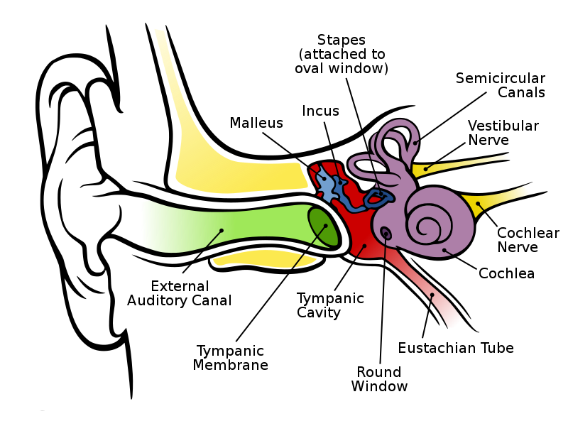
Anatomy of the ear.
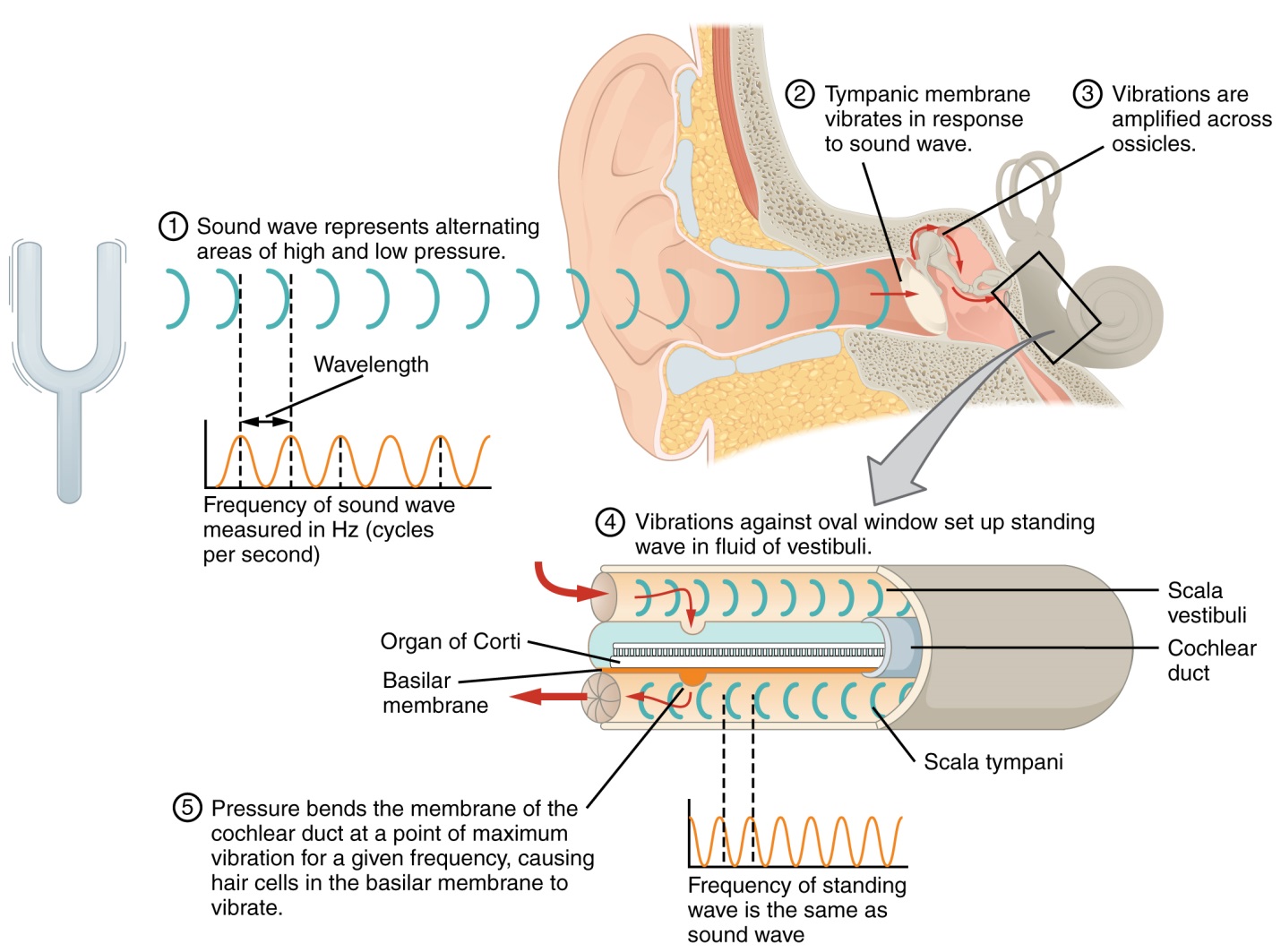
Events involved in audition (hearing).
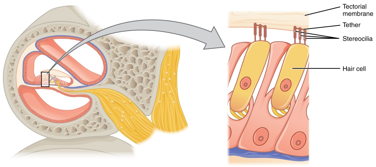
Organ of Corti inside cochlea, and magnified view of hair cells.
Perception of Sound
Sound, as defined in physics, is a vibration that propagates as a wave of pressure through a gas, liquid, or solid. Sounds are perceived differently based on characteristics of these waves. One feature of sound waves that affects their perception is their amplitude. The higher the amplitude of a sound wave, the louder that sound will be perceived. There is also a relationship between a sound wave’s frequency and its perceived pitch. The frequency of a sound wave has an inverse relationship with the wavelength of that wave (in other words, the longer the wavelength, the lower the frequency; conversely, the shorter the wavelength, the higher the frequency of that wave). The frequency of sound waves is usually measured units called Hertz, which are defined as cycles per second. You have been provided with several tuning forks, each of which is stamped with its vibrational frequency in Hertz. Take one of the provided tuning forks, and note its frequency. Strike the tuning fork on the provided rubber wedge, and note the sound produced. Repeat this for the other tuning forks. Did you notice the relationship between the frequency and pitch of each tuning fork? Answer the question regarding this on the worksheet at the end of this lab exercise.
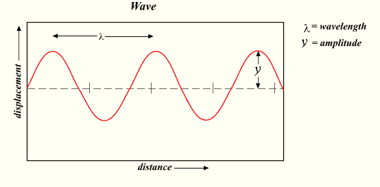
Characteristics of waves.
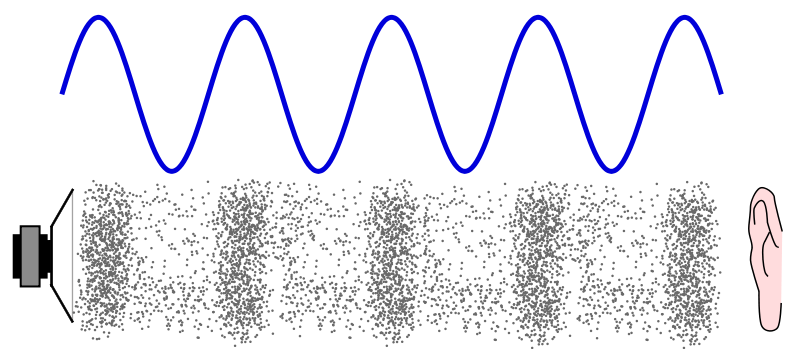
Diagram showing physical manifestation of a sound wave through air from a speaker to a human ear
Locating Sound
- Sounds are located using the combined perception of those sounds by both ears. Differences in hearing between the ears can result in an incorrect determination for the location between sounds. In this exercise, you and your partner will explore your abilities to determine the location of sounds. Follow the directions below to conduct this activity:
- Have your partner be seated, with eyes closed.
- Strike the provided tuning fork with the lowest frequency at one of the following locations relative to your partner’s head (use a random order for these):
a. Directly below and behind the head: ______________________________________
b. Directly behind the head: ______________________________________
c. Directly above the head: ______________________________________
d. Directly in front of the face: ______________________________________
e. To the right side of the head: ______________________________________
f. To the left side of the head: ______________________________________
4. Ask your partner to give the exact location of each sound, and record their responses in the blanks above.
5. Repeat the process using the provided tuning fork with the highest frequency, and record your partner’s responses below:
a.Directly below and behind the head: ______________________________________
b. Directly behind the head: ______________________________________
c. Directly above the head: ______________________________________
d. Directly in front of the face: ______________________________________
e. To the right side of the head: ______________________________________
f. To the left side of the head: ______________________________________
6. Switch roles with your partner, with yourself trying to determine the location of sound as above.
7. Answer the questions regarding this activity on the worksheet at the end of this lab exercise.
The inner ear also contains several organs collectively referred to as the vestibular system, shown in the figure on the following page. The organs of the vestibular system include the vestibule, the utricle, the saccule, and three semicircular canals. Each of these organs contains hair cells, similar to the cochlea, but are not involved with hearing. Instead, these organs are involved with equilibrium, balance, and detection of positional information and directional acceleration. The saccule and utricle sit below a gelatinous layer of the vestibule, with the cilia of their hair cells projected into the gelatin. Inside the gelatin layer are crystals of calcium carbonate called otoliths (which literally means “ear stones”). Changes in the angular position of the head causes these crystals to shift, bending the cilia of the hair cells in the utricle and saccule. The bending of the hair cells stimulates neurons, which carry this signal information to the brain via the vestibular nerve.
The semicircular canals are fluid-filled tube-like structures that also contain hair cells. These canals are involved in the detection of angular acceleration and deceleration from rotation. When turning your head, this shifts fluid in these canals, which also bends the cilia of hair cells inside the canals, and signals from the canals are sent to the brain for processing. When movement in a particular direction is accelerated or decelerated, this also causes movement of fluid in these canals. One way to think about this is if you are standing still and holding a glass of water, and suddenly begin walking quickly forward, some of the water may splash backwards onto your hand. Then, if you suddenly stop walking, the water may splash forward. In this way, the semicircular canals are responsive to changes in velocity. This information is also relayed through the vestibular nerve to the brain for processing.
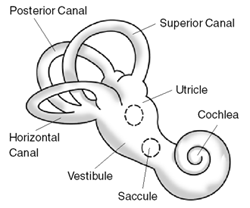
Organs of the vestibular system of the inner ear.
Illustration of Post-rotatory Nystagmus
In this activity, you will examine nystagmus, which is the movement of the eyes in response to stimulation of hair cells in the semicircular canals. Nystagmus displays two component motions, called the slow phase and the fast phase. When the head is rotated, the eyes move slowly in the opposite direction, then quickly back to the other side, and then begins the slow movement again. Using the steps below, you will induce post-rotatory nystagmus, which is the result of repeated rotational movement, followed by cessation (stopping) of that movement. To illustrate post-rotatory nystagmus, follow the steps below:
- Get your partner to sit in his or her chair, and raise his/her feet off the floor.
- Your partner should then bend their head forward at an angle of about 30 degrees.
- Rotate your partner in their chair to the right (clockwise) for about 20 seconds at a rate of one turn every two seconds. (Be careful not to fling your partner out of their chair!)
- Suddenly stop the rotation, and quickly observe the movement of your partner’s eyes as soon as the rotation stops, noting the direction of movement of the slow and fast phases of nystagmus.
- Repeat Steps 1-4, only this time rotating your partner to the left (counter-clockwise).
- Answer the question on the worksheet at the end of this lab exercise.
- Repeat, switching roles with your partner so that they may observe nystagmus of your eyes.
The Chemical Senses: Smell and Taste
Both smell and taste are chemical senses, in which chemical stimuli are received by chemoreceptors (in the nose in the case of smell, and on the tongue in the case of taste). In the following activities, you will explore your own senses of smell and taste, as well as familiarize yourself with the anatomy of organs involved in each of these senses, as well as the physiology of how chemical stimuli are detected and processed.
Sense of Smell
Olfaction is the technical term used to describe the sense of smell. In the superior (upper portion) nasal cavity, there are multiple olfactory receptor cells, which are hair cells with cilia, similar to those seen in the cochlea and the vestibular apparatus. As molecules in the air are inhaled, they pass over the olfactory epithelial tissue and become dissolved in the mucus coating the surface of that tissue. These molecules then bind to proteins, which help transport them to the olfactory dendrites. Once these odorant molecules (and proteins to which they are bound) reach the olfactory dendrites, they bind to receptor proteins in the dendrites’ cell membranes. The binding of these molecules to the olfactory dendrites is then converted to an electrical signal, which is carried to the olfactory bulb on the inferior (lower) surface of the brain. This information is then carried via the olfactory tract for processing by various parts of the brain, including the olfactory cortex, hippocampus, amygdala, and hypothalamus. Many of these parts of the brain are also part of the limbic system, which is associated with emotions and memory. This is one reason that certain smells often evoke certain memories associated with those particular scents.
Humans have a total of about 40 million olfactory receptors, each of about 350 different subtypes. Stimulation of different olfactory receptors in various combinations allow us to distinguish about 10,000 different odors. Many other mammals have an even greater sense of smell. For example, mice have about 1,300 different types of olfactory receptors, almost 4 times as many as humans, and so are probably able to distinguish many more odors than humans. Dogs also have LOTS more olfactory receptors than humans. Though the number of olfactory receptors in dogs varies among individuals and different breeds, just as an example, German shepherds have about 2 BILLION olfactory receptors (individual receptor cells, not subtypes), which is four times the number of total olfactory receptors in humans!
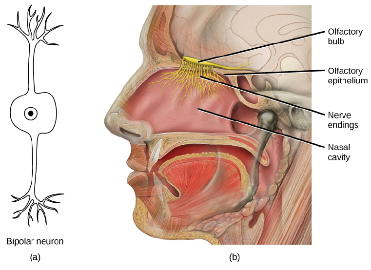
Illustration of (a) bipolar neurons found in the nasal cavity, and (b) olfactory epithelial tissue and the olfactory bulb.
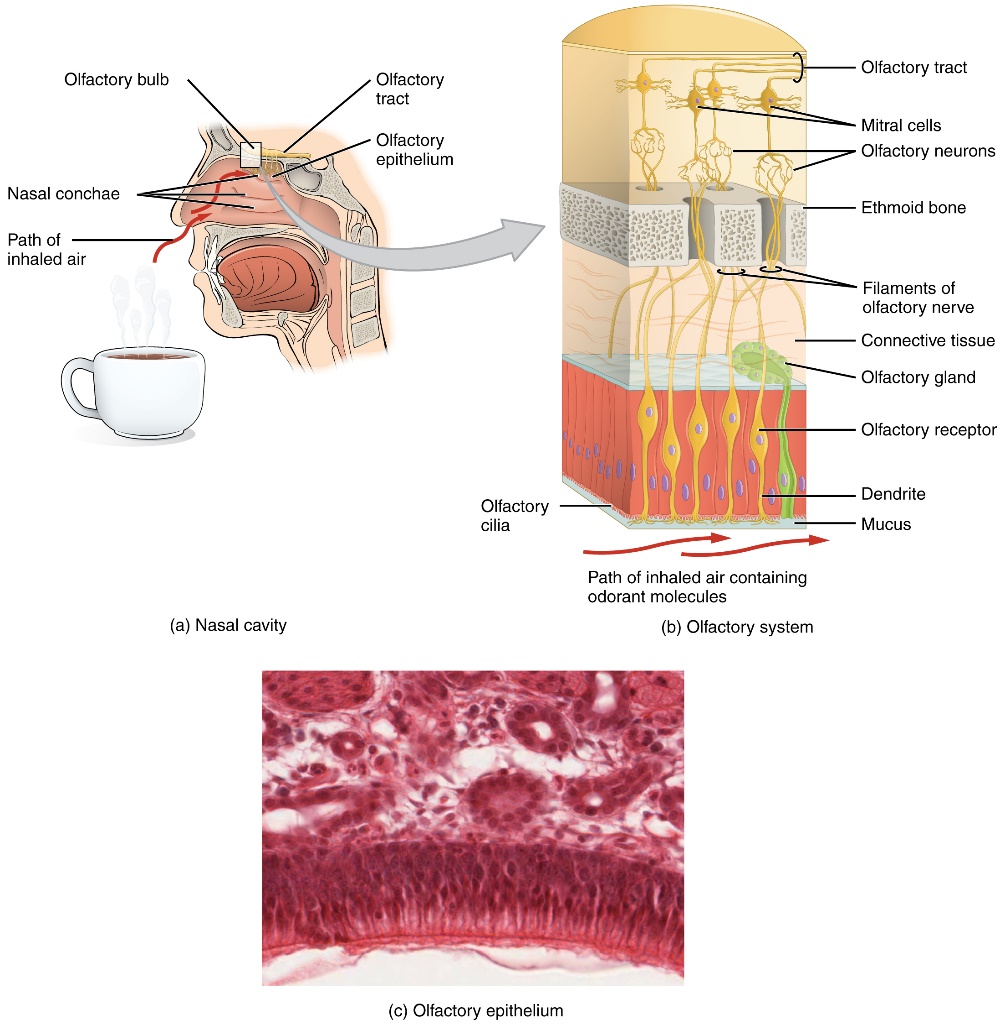
The olfactory system
To test your olfactory abilities, you will use several vials containing artificial and natural scents. Each table has been supplied with 10 of these vials, numbered 1-10. One pair of students at each table has a basket with vials 1-5, and the other pair of students should have vials 6-10. For this exercise, you will need to smell all 10 vials, so share with your tablemates as necessary. For each vial, you should unscrew the lid and smell the contents. Though not necessary, you may wish to close your eyes to reduce visual stimuli and focus more on your sense of smell. After smelling each vial, record what you think the scent in that vial is supposed to be in the table on the worksheet at the end of this lab exercise. After smelling each vial and recording what scent you think is in each vial, your instructor will provide you with the actual scents to compare to your guesses, and to also record in the table.
Sense of Taste
Like olfaction (the sense of smell), gustation (the sense of taste) is also a chemical sense, involving chemoreceptor cells that respond to chemical stimuli, convert that information to electrical signals that is relayed to the brain for interpretation. Though smell and taste are often perceived as completely separate senses, the two senses do work together to form impressions of perceived flavors. You may have noticed, for example, that your sensation of flavor is dulled somewhat if you have a stuffy nose.
The primary organ of taste is the taste bud, which is a cluster of taste cells. Taste buds are found on bumps, called papillae, on the tongue. Look at the figure on the following page, and notice that there are several different types of papillae on the tongue, and each type differs in the numbers of taste buds they possess. For example, the filiform papillae, found all over the tongue, do not have any taste cells. The fungiform papillae, which are mainly located on the anterior (front) two thirds of the tongue, contain 1-8 taste buds each, as well as pressure and temperature receptors. The circumvallate papillae, which are quite large, and form an upside-down “V” near the rear of the tongue contain 100-250 taste buds. Finally, the foliate papillae, which are found in parallel folds along the edges and back of the tongue, contain approximately 1,300 taste buds each. Taste bud cells are replaced about every 10 to 14 days.
When eating, molecules in food are dissolved in saliva, and bind with and stimulate taste hairs on the tips of taste bud cells. Most of the taste receptors on the tongue are found on the outer edge and front of the tongue. There are different types of taste receptors for different flavors. Humans perceive five main primary tastes, and each of these different types of tastes has one corresponding type of receptor. The five primary tastes humans can detect include salty, sour, sweet, bitter, and umami. Salty foods contain sodium chloride, and ingesting them dissolves the sodium chloride into sodium and chloride ions. The sodium ions directly enter into taste neurons, exciting them. Sour tasting foods contain acids. When acids bind to receptors, that triggers hydrogen ion channels to open, allowing hydrogen ions into the receptors, triggering them. Sweet, bitter, and umami tastes require a special kind of receptor (called a G-protein coupled receptor, or GPCR), which uses a protein (a G protein) that acts as a molecular switch inside cells. The sense of taste has a tendency to decline with age, often dramatically beginning around age 50.
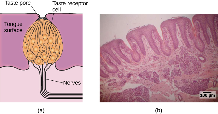
(a): Schematic drawing of a taste bud.
(b): micrograph of human tongue tissue.
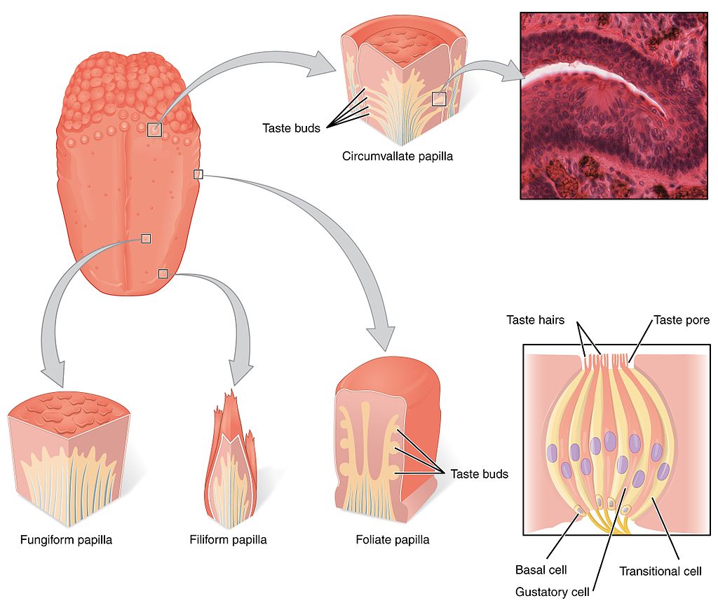
Illustration of the various types of papillae on the tongue, as well as a magnified view of a taste bud.
In the following exercise, you will examine your own sense of taste, as well as learn something about the microscopic anatomy of your own tongue. For this exercise, you have been provided with 4 vials of paper strips. One vial of these strips contains “Control” paper, which is just plain paper. The other vials contain strips of paper which are impregnated with various chemicals, including Phenylthiocarbamide (PTC), thiourea, and sodium benzoate. The interesting thing about each of these chemicals is that some people vary in terms of whether they are able to taste those compounds. Interestingly, the ability to taste each of these compounds is genetic, as the genes involved in whether individuals do/do not taste these compounds are genes that affect the expression of specific receptor proteins in taste buds.
For example, the ability to taste PTC (detecting it as a bitter flavor) is due to a dominant allele. Non-tasters thus have two recessive alleles coding for the receptor protein in question. Individuals that are homozygous dominant (have two dominant alleles) may perceive the taste of PTC to be more bitter than heterozygotes (individuals with one dominant and one recessive allele), though heterozygotes are able to taste PTC as bitter. In a group of studies, individuals that were tasters of PTC tended to avoid broccoli and grapefruit juice, and tend to find green vegetables quite bitter.
Thiourea is another compound somewhat similar to PTC, but the ability to taste it is inherited independently. Thus, the ability to taste PTC does not mean that PTC tasters will also be able to taste thiourea, and vice versa. To tasters, thiourea also tends to have a very bitter taste.
Sodium benzoate is commonly used as a preservative in many foods, but in very low concentrations. However, some people are very sensitive to its taste. Perceptions of the taste of sodium benzoate (in tasters) varies, with some perceiving it as sweet, some as salty, some as bitter, and some as sour. The ability to taste sodium benzoate is also inherited independently of the ability to taste PTC, but the interactions to sensitivity to each of these molecules has been shown to influence people’s reactions to different foods. For example, PTC tasters that taste sodium benzoate as salty tend to like sauerkraut, buttermilk, turnips, and spinach more than the average person, and PTC tasters that taste sodium benzoate as bitter tend to like those foods less than the average person.
- Using the provided vials of control, PTC, thiourea, and sodium benzoate paper, follow the directions below:
- Obtain a piece of “Control Paper”. This strip of paper is nothing more than paper, and will be used for a basis for comparison to the other strips.
- Place the piece of control paper in your mouth. You should notice no strong flavor, other than just the bland flavor of the paper itself. Try to remember the general baseline flavor of this paper.
- Remove the control paper from your mouth and throw it away.
- Next, take a piece of PTC paper, and put it on your tongue. If you do not immediately detect a noticeable flavor, briefly chew the strip of PTC paper. If you do not notice any flavor at all, you are a non-taster for PTC.
- Remove the PTC paper from your mouth (do not swallow it), and throw it away.
- Repeat steps 5 & 6 for both the thiourea paper and the sodium benzoate paper.
- Record your observations of your perceived taste/lack of taste of each compound in the table on the worksheet at the end of this lab exercise.
- ______________________________________________________________________________
Image Credits:
Accommodation By Pearson Scott Foresman – Archives of Pearson Scott Foresman, donated to the Wikimedia Foundation, Public Domain, https://commons.wikimedia.org/w/index.php?curid=3606348
Near Point Chart By http://www.ssc.education.ed.ac.uk/courses/outreach/dublini.html
Astigmatism By The original uploader was Tallfred at English Wikipedia – Originally from en.wikipedia; description page is/was here., BSD, https://commons.wikimedia.org/w/index.php?curid=3098293
Astigmatism By BruceBlaus (Own work) [CC BY-SA 4.0 (https://creativecommons.org/licenses/by-sa/4.0)], via Wikimedia Commons
Anatomy of the ear: Chittka L. Brockmann – Perception Space—The Final Frontier, A PLoS Biology Vol. 3, No. 4, e137 doi:10.1371/journal.pbio.0030137 (Fig. 1A/Large version), vectorised by Inductiveload; Licensed under Creative Commons Attribution 2.5 Generic.
Audition: https://archive.cnx.org/contents/238b840d-2428-4d16-bcd3-ab481a44522a@1/derived-copy-of-sensory-perception
Organ of Corti: https://archive.cnx.org/contents/238b840d-2428-4d16-bcd3-ab481a44522a@1/derived-copy-of-sensory-perception
Sound wave diagram: Wikimedia Commons; author Pluke; licensed under Creative Commons – CC0 1.0 Universal
Vestibular system: http://www.nidcd.nih.gov/health/balance/balance_disorders.asp
Olfactory neurons, epithelial tissue, and olfactory bulbs: modification of work by Patrick J. Lynch, medical illustrator; C. Carl Jaffe, MD, cardiologist
Olfactory system: https://archive.cnx.org/contents/13be3d63-b803-4d7b-9265-14da1c5585ae@1/sensory-perception
Closeup of taste bud: Jonas Töle; released to public domain; Micrograph of tongue tissue: https://archive.cnx.org/contents/370f4538-a2ec-4bf7-a3f4-c00b15548c04@5/the-other-senses
Tongue & papillae: https://legacy-staging1.cnx.org/content/m10555/latest/?collection=col10044/latest (modification of work by Vincent Rizzo).
BI 102 Lab Worksheet: Senses Name _________________________________ Section _______
Visual acuity:
Left eye:20/___ Right eye:20/__
Astigmatism:
Left eye: ______________ (yes/no) Right eye: ______________ (yes/no)
Accommodation:table 30.2
Left eye: ______ cm “Age” of your L eye: _____
Right eye: ______ cm “Age” of your R eye: _____
Blind spot:
Left eye: ______ cm Right eye: ______ cm
Color Blindness Score: _______ (count # of plates you cannot read)
Superimposition
Describe what you saw during the superimposition exercise.
Afterimages:
Describe what you saw during the afterimages exercise using the strip of green paper.
Describe what you saw during the afterimages exercise using the strip of orange paper.
2-point discrimination (touch):
Forearm: ___ mm Back of neck: ___ mm Index finger: ___ mm Back of hand: ____ mm
Which area contains the greatest density of touch receptors? Why do you think this is the case?
Sense of Heat and Cold:
Describe the sensations experienced in your left and right hands during this exercise.
The Ear, Hearing, and Equilibrium:
Number the following structures below in the order in which sound waves/vibrational pass through them.
_____ Auditory canal
_____ Cochlea
_____ Cochlear nerve
_____ Incus
_____ Malleus
_____ Oval window
_____ Pinna
_____ Stapes
_____ Tympanic membrane
Perception of Sound:
What is the relationship between the frequency of a sound and its perceived pitch?
Locating Sound:
Was it more difficult for you to accurately locate sounds with lower or higher pitches?
Illustration of Post-rotatory Nystagmus:
Describe the direction of eye movements during the slow and fast phases of nystagmus below:
After rotating clockwise: Direction of slow phase: __________ Direction of fast phase: __________
After rotating counter-clockwise: Direction of slow phase: _______ Direction of fast phase: _______
Sense of Smell:
|
Vial # |
What you think is the scent |
The actual scent |
|
|
|
|
|
|
|
|
|
|
|
|
|
|
|
|
|
|
|
|
|
|
|
|
|
|
|
|
|
|
|
|
|
|
|
|
|
|
|
|
Sense of Taste:
|
Type of Paper |
Describe taste (no taste, sweet, sour, salty, or bitter) |
|
Control |
No taste |
|
Phenylthiocarbamide (PTC) |
|
|
Thiourea |
|
|
Sodium benzoate |
|
