5 Chapter 5
LAB 5
Homeostasis
Prepared by Dr. Jeff Ray, Dept. of Biology, UNA
OBJECTIVES
After completing these laboratory activities, you should understand / be able to:
- Homeostasis as the central theme of physiology and the importance of the liver, kidneys, and lungs in this process.
- The liver’s role in maintaining blood glucose homeostasis and why the serum from blood vessels in proximity to the liver will have differing amounts of glucose immediately after eating a meal versus after fasting for 6 hours.
- The kidney’s role in maintaining homeostasis, explain the 4 basic steps of kidney function, and name substances that should/should not be in the urine.
- The results of the urinalysis and identify the abnormal values in the patient.
- The lungs’ role in homeostasis and know what vital capacity represents.
Introduction
Homeostasis is the central theme of physiology and refers to the dynamic equilibrium of the body’s internal environment. Parameters like body temperature, blood glucose levels, heart rate, and other values are constantly fluctuating above and below a set point (varying within tolerable ranges). To maintain values near the set point (e.g. 98.6° F for body temperature), feedback systems/loops in the body use receptors to detect change, a control center to process the information, and an effector to carry out the change in a feedback loop (pathway of the loop is receptor control center effector). Receptors include chemoreceptors (that detect molecules like CO2), a control center (normally the brain, especially the hypothalamus); and an effector (usually a muscle or gland). Homeostasis is primarily maintained by negative feedback. Negative feedback involves adjustments that oppose the initial change (i.e. cause change in the opposite direction). Positive feedback is less common and often irreversible; examples include digestion of proteins, blood clotting, and action potentials in neurons. All organs system contribute to homeostasis, but the three particularly important organs within the digestive, respiratory, and urinary systems are the liver, lungs, and kidneys, which exchange materials with the blood.
Today’s lab, activities will illustrate the central roles played by the liver (maintain blood glucose levels), kidneys (filter wastes from blood), and lungs (exchange O2/CO2) in maintaining homeostasis.
Liver
The liver is the largest organ in the abdominal cavity, averaging three pounds in the adult. It is located in the upper right quadrant, immediately beneath the diaphragm and on top of the stomach. The liver has a variety of roles (500+ known) in the body including: (1) store/release vitamins and minerals, (2) produce bile, (3) produce blood proteins, and (4) detoxify substances like alcohol.
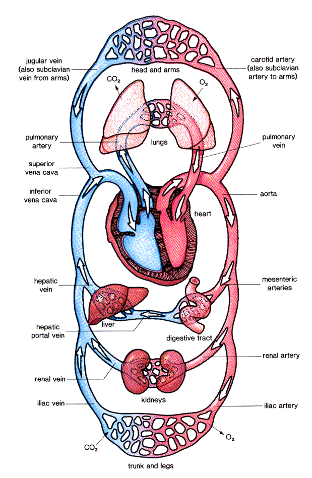
One major role of the liver is to maintain blood glucose homeostasis and it is ideally positioned to serve this role, due to its proximity to the digestive tract. There are several major blood vessels associated with this activity. Carrying blood from the aorta to the digestive system is the mesenteric artery. In the digestive tract, nutrients are gathered and funneled into the hepatic portal vein which leads to the liver; exiting from the liver is the hepatic vein.
The pathway is (mesenteric artery→ digestive tract→ hepatic portal vein→ liver→ hepatic vein).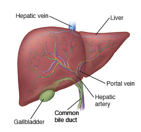
Blood glucose homeostasis is a negative feedback mechanism. Whether one has recently eaten or not, cells require a constant supply of glucose (sugar), which is the direct fuel to produce ATP. As a result, the liver must keep the glucose level in blood at around 0.1% by storing excess glucose (as glycogen) and releasing glucose as needed.
Another organ, the pancreas, actually monitors glucose levels and signals the liver with hormones. After eating, blood glucose levels rise. This increase is detected by the pancreas, which in response secretes insulin. Insulin travels through the bloodstream and binds to cells in the liver signaling the cells to absorb glucose and store it as glycogen. While fasting, blood glucose levels begin to drop, this reduction is detected by the pancreas, which secretes glucagon, a hormone that causes the liver to release glucose, thereby maintaining blood glucose levels near 0.1%. (insulin signal: store glucose; glucagon signal: release glucose).
Basic Instructions: Blood Glucose Homeostasis
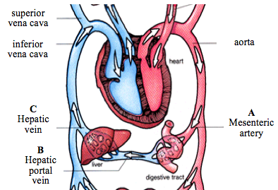
In the following exercise, you will conduct a simulation of blood glucose levels in three blood vessels (see figure).
A = Mesenteric artery (before digestive tract)
B = Hepatic portal vein (b/w digestive–liver)
C = Hepatic vein (after liver)
You will observe differences in blood glucose levels in each of these vessels under two simulated conditions:
1 = immediately after eating a meal
2 = after fasting for 6 hours
* Solutions labeled A1, B1 & C1 simulate blood glucose levels immediately after eating.
* Solutions labeled A2, B2 & C2 simulate blood glucose levels after fasting for 6 hours.
Follow the directions carefully. Check each step after completing it:
____ Prepare a boiling water bath: fill a 400 mL beaker about 2/3 full with tap water; place it on your
hot plate. Do NOT let all of the H2O evaporate or the beaker may break- add H2O as needed.
____ Using a Sharpie, label 6 test tubes “A1”, “B1”, “C1”, “A2”, “B2”, and “C2”.
____ To each test tube add 2 pipets of the appropriate solution. Use the labeled pipets ONLY for their
intended solutions to avoid cross-contamination of solutions.
____ Add 3 pipets of Benedict’s solution* to each of your test tubes.
(*Benedict’s solution tests for the presence of glucose. After boiling, Benedict’s solution will change from blue if glucose is present (blue/cloudy= no/low glucose, yellow/orange= moderate glucose, red= high glucose).
____ After your water bath is boiling, add tubes A1, B1, and C1 to the boiling bath AT THE SAME
TIME,
and WATCH CLOSELY for the order of change.
____ Record the order in which the solutions in the tubes changed color in the table below.
A FASTER COLOR CHANGE INDICATES A HIGHER CONCENTRATION OF GLUCOSE.
____ If no color change is observed in the last tube after 2 minutes of boiling, you may stop the procedure.
Blood Glucose Levels After Eating (Time 1)
|
Test tubes (in order of change) $ |
|
Blood vessel associated with the tube # |
|
1st |
|
|
|
2nd |
|
|
|
Last |
|
|
$
answer will be A1, B1 or C1 # answer will be mesenteric artery, hepatic portal vein or hepatic vein
* Which blood vessel contains the most glucose after eating a meal? _____________________________
*Why did the hepatic vein contain less glucose than the hepatic portal vein after just eating a meal? ________________________________________________________________
____ Remove tubes A1, B1, and C1 from the boiling water bath.
____ Add tubes A2, B2, and C2 to the boiling bath AT THE SAME TIME, and WATCH CLOSELY.
____ Record the order in which the solutions in the tubes change color in the table below.
____ If no color change is observed in the last tube after 3 minutes of boiling, you may stop the procedure.
____ *Clean your station: Turn off your hot plate. Dump liquids from tubes down the drain with water running, place used test tubes in tub. Return all materials to their starting location. Wipe off your table.
Blood Glucose Levels After Fasting for 6 hours (Time 2)
|
Test tubes (in order of change) $ |
|
Blood vessel associated with the tube # |
|
1st |
|
|
|
2nd |
|
|
|
Last |
|
|
$
answer will be A2, B2 or C2 # answer will be mesenteric artery, hepatic portal vein or hepatic vein
*Which blood vessel contains the most glucose after fasting for ~6 hours? ________________________
*Why did the hepatic vein contain more glucose than the hepatic portal vein after fasting? ___________________________________________________________________________________
*So, the role of the liver in maintaining blood glucose homeostasis is to store extra glucose (as glycogen) after eating and release glucose during fasting (glucose fuels our cell’s activities 24/7)*
Kidneys
Kidneys are fist-sized organs located along the dorsal abdominal wall, behind the intestines. Their basic function is to filter metabolic wastes and toxins from the blood to produce urine. Specifically, kidneys excrete nitrogen by-products and regulate blood volume, blood pressure, and pH. Large substance like red blood cells, white blood cells, and proteins do not normally leave blood vessels and get filtered by the kidneys, these should not be found in the urine, but normally stay in the blood. Small molecules move into the kidneys filtering units called nephrons, and either must be excreted by the kidneys in the urine or be recovered for use in the body. Urea is a small molecule and waste product of protein metabolism that must be excreted. Other substances like glucose move into the kidneys, but are mostly recovered for use by the body, although excess (abnormal) amounts will be disposed of in urine. Thus, whatever is/is not in urine gives insight into kidney function. Learning the structure and function of the nephron will help in understanding urinalysis results.
Kidneys collectively filter 180 liters of blood plasma daily to produce about 1.5 liters of urine/day.
Although complex, the basic steps of nephron functioning are 1) filtration, 2) reabsorption, 3) secretion, and 4) excretion. Filtration is the movement of water and solutes from plasma into the nephron and is primarily driven by blood pressure pushing into the first part of the nephron (glomerulus) that is very permeable. Once in the nephron, this liquid becomes the filtrate. The filtrate continues through the tubules and is reabsorbed. Reabsorption is the movement of water and solutes back into the network of capillaries surrounding the tubule. Substances that are waste products or in excess amounts move back into the tubules via secretion. Secretion is essential to regulate blood volume, pH and electrolyte levels. Excretion eliminates substances like urea in the filtrate, which is now “urine”.
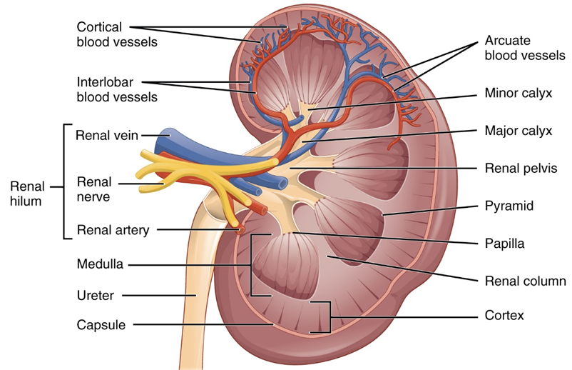
Kidney & blood vessels; ureter empties to the bladder.
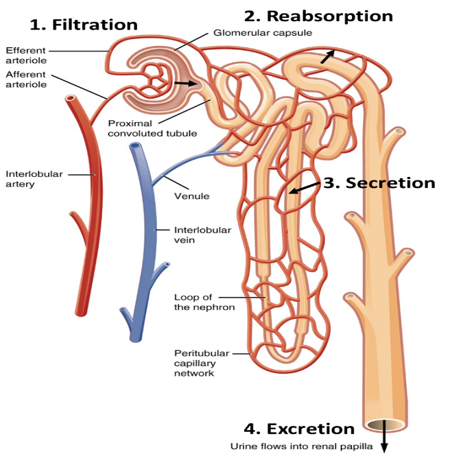 Nephron, the functional unit of the kidney showing the 4 steps of cleansing the blood.
* What is the kidneys’ job in maintaining homeostasis? _______________________________________
Urinalysis
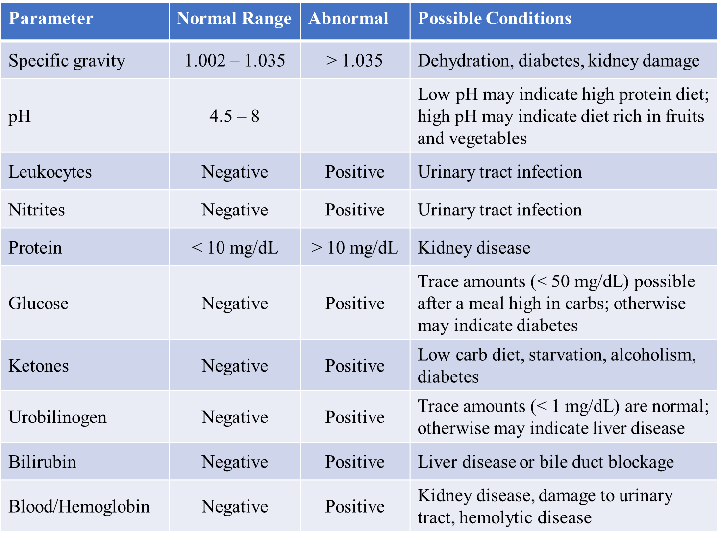
A urinalysis is a non-invasive procedure that gives insight into the basic health and functioning of kidneys and other organs, including the liver. You will conduct the same urinalysis that is regularly performed in a doctor’s office using artificial urine. This test is different from those given to job applicants, which screens for illegal drugs in the body. A basic urinalysis uses urine test strips to measure 10-12 values that either: (1) should be within a certain range (specific gravity, pH) or (2) detects substances that should not be in the urine (e.g. leukocytes- white blood cells), because they normally remain in the blood since they are too large and do not pass into the nephrons of the kidneys. Chemstrip 10’s will be used to look at 10 different parameters of the urine. The color and odor of urine also give insight into body conditions (like dehydration) as does the microscopic examination of substances in urine. Abnormal values may be temporary and do not necessarily indicate any long-term problems, but a doctor might request additional tests as a follow-up to confirm any values outside of the normal range.
Patient #1 reports that they are often thirsty, but also urinate frequently. Patient #1 eats a normal diet of approximately 2,000 calories, but has experienced weight loss over the past 6 months. The individual also feels tired and run down. What condition is likely affecting patient #1? A urinalysis may help diagnose their disease. Follow the directions below and record your results.
Basic Instructions: Urinalysis
____ Obtain a Chemstrip 10 urinalysis test strip. Lay this strip on top of two paper towels on your lab table.
____ Using the pipet, place 1 drop of patient #1’s simulated urine on each colored square on the test strip, do not let urine from separate squares run together. Holding the test strip by the handle, turn the strip on its edge and gently tap any excess urine onto the paper towel.
____ Within 1 minute, compare the results on the test strips to the scale on the side of the test strip vial (read handle side up). You may also try the automated urinalysis reader in the back of the room (these are used in Dr.’s offices). Record your results below.
____ *Do your own urinalysis – see professor for additional instructions*
____ Clean up your station: throw away used paper towels and test strips. If you did your own urinalysis, put the urine cup where your instructor tells you, do not leave it at your station.
URINALYSIS TESTING – circle any abnormal values
Patient Values (normal) Your Values (normal)
1. _____ Specific gravity (1.005-1.035) 1. _____ Specific gravity (1.005-1.035)
2. _____ pH (=acidity; varies) 2. _____ pH (=acidity;varies)
3. _____ Leukocytes (=WBC’s; negative) 3. _____ Leukocytes (=WBC’s; negative)
4. _____ Nitrite (negative) 4. _____ Nitrite (negative)
5. _____ Protein (negative) 5. _____ Protein (negative)
6. _____ Glucose (<50) 6. _____ Glucose (<50)
7. _____ Ketones (negative) 7. _____ Ketones (negative)
8. _____ Urobilinogen (<1) 8. _____ Urobilinogen (<1)
9. _____ Bilirubin (negative) 9. _____ Bilirubin (negative)
10. _____ Blood (negative) 10. _____ Blood (negative)
Diagnosis: __________________ Diagnosis: __________________
* The 3 values for the patient that were abnormal were:
a. _________* Low (=acidic), Moderate (neutral=7.0) or High (basic, >7)
b. _________Low, Moderate or High
c. _________ Low, Moderate or High
*What is the diagnosis of your patient (what condition)? ________________________________
Lungs
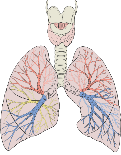
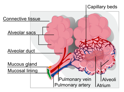
Lungs are located in the pleural cavities (overall within the thoracic cavity) and are lateral to the heart. The right lung has three lobes and is larger than the left, which has two lobes; the lungs collectively weigh about 3 pounds. Lungs exchange gases with the blood (O2 in / CO2 out) via simple diffusion- no cellular energy is required. The lungs contain approximately 1,500 miles of airways and 300 to 500 million alveoli, which appear as grape clusters. Gas exchange occurs at alveoli, which contain single-layered flat cells (simple squamous epithelium) that maximize surface area exchange.
Respiration
The physical process of breathing, respiration, includes inhalation and exhalation. The active part of breathing is inhalation and involves a dome-shaped breathing muscle, the diaphragm.
Different measures of lung function are made based upon volumes of air (see chart below), but the most commonly reported measure is vital capacity (see figure next page), which is the maximum amount of air a person can expel from the lungs after a maximum inhalation. Vital capacity is measured with a spirometer (in liters or milliliters) and may aid in a diagnosis of underlying lung disease if vital capacity is reduced.
Basic Instructions: Lung Volume
Clean the spirometer with an alcohol swab before & after use. Put a clean, disposable mouthpiece on the spirometer. Set spirometer dial to zero by turning the silver ring on top. While standing, take a full breath OUT. Breathe in fully, then place your mouth on the disposable mouthpiece & blow all air from your lungs into spirometer. Do NOT breathe IN with spirometer to your lips. You may also try the lung volume bags on the front table.
Record your vital capacity in Liters three separate times, then calculate the average of the three values.
1. ___________ Liters 2. ___________ Liters 3. ___________ Liters
Average vital capacity ___________ Liters
*Average volumes in adults are 2-4 liters: females & 3-5 liters: males.
______________________________________________________________________________
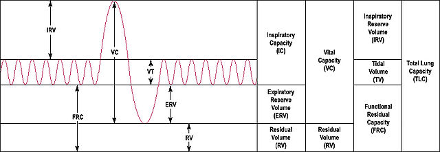
Lung Measures including Vital Capacity
Basic Instructions: Body Composition Monitor
Enter your information into the handheld monitor following the onscreen prompts. Record your information in the blanks below. Determine your BMI based on the chart that is with the body composition monitor with the understanding that BMI does NOT fully account for differences in body types in determining obesity.
1. _______ Height (inches) 2. _______ Weight (pounds) 3. _______ Age (years)
4. _______ Male or Female 5. _______ Normal or Athletic
A. _______ Body Mass Index (BMI) B. _______ Body Fat Percentage (%)
Concept Questions – consult your textbook if needed
How do the kidneys respond when the blood pressure and volume are too high? Too low?
_________________________________________________________________________________
_________________________________________________________________________________
List the steps in urine formation and define each step
1.______________________________________ 2. _________________________________________
3. _____________________________________ 4. __________________________________________
With regard to urine formation, name a substance found in both the filtrate and the urine.
_________________________________________________________________________________
With regard to urine formation, name a substance found in the filtrate and not in the urine.
_________________________________________________________________________________
Image Credits:
Liver by http://www.stanfordchildrens.org/en/topic/default?id=how-the-liver-works-90-P02006
Systemic Circulation By OpenStax College – Anatomy & Physiology, Connexions Web site. http://cnx.org/content/col11496/1.6/, Jun 19, 2013., CC BY 3.0, https://commons.wikimedia.org/w/index.php?curid=30148241
Kidneys and Nephron
https://cnx.org/contents/FPtK1zmh@12.8:7l9EIHui@7/Gross-Anatomy-of-the-Kidney
Alveoli By LadyofHats – self-made (extracted from Image: Respiratory system complete.svg) (duplicate of Image:Respiratory system complete en.svg), Public Domain, https://commons.wikimedia.org/w/index.php?curid=3222341
Lungs By Patrick J. Lynch, medical illustrator – Patrick J. Lynch, medical illustrator, CC BY 2.5, https://commons.wikimedia.org/w/index.php?curid=1496626
Lung volumes By Original uploader was Vihsadas at en.wikipedia – Transferred from en.wikipedia, Public Domain, https://commons.wikimedia.org/w/index.php?curid=4145884
BI 102 Lab Worksheet: Homeostasis Name ___________________________ Section _______
1. What is the liver’s job in maintaining blood glucose homeostasis?
2. Sketch the locations of the mesenteric artery, hepatic portal vein and hepatic vein in relation to the digestive system and liver.
3. What is the kidneys’ job in maintaining homeostasis?
4-6. The 3 values for Patient #1 that were abnormal were:
a. _________* Low (=acidic), Moderate (neutral=7.0) or High (basic, >7)
b. __________Low, Moderate or High
c. __________ Low, Moderate or High
*by itself, not a cause for concern, may be abnormal due to mild dehydration or overhydration
7. What is the diagnosis of your Patient #1 (what condition)?
8. Which value was High due to the breakdown (metabolism) of fats?
a. glucoseb. ketonesc. bilirubin
9. Name 2 of the substances tested for which you would NOT expect to find in a normal urinalysis.
10.What is the lungs’ job in maintaining homeostasis?
11. Avg. volumes in adults are ___-___ liters: females & ___-___ liters: males. How did your values compare?
