2 Chapter 2
LAB 2
Musculoskeletal System
Prepared by Dr. Jeff Ray, Dept. of Biology, UNA
OBJECTIVES
After completing these laboratory activities, you should understand/be able to:
- The basic structure, function, and total number bones and muscles in the skeletal and muscular systems, including the axial and appendicular divisions.
- The tissues of these systems, including bone, hyaline cartilage, and skeletal muscle.
- Identify the specific bones (including the vertebral regions) and muscles as specified by your instructor.
- The types of joints, muscle movements, and basics of muscle contraction.
Introduction
The muscular and skeletal systems collectively make up a large percentage of body mass and are studied together due to the interdependence and interaction between these organ systems in supporting and moving the body. Joints are at the interface of where bones meet and are moved by muscles. The skeletal system provides a scaffolding for holding up the body and assisting movements. It functions include support, movement, protection, blood cell production, storage of minerals, and endocrine regulation. The human adult normally has 206 bones.
The muscular system functions in moving bones, provides posture and support, protects internal organs, and generates heat. The more than 600 skeletal muscles are composed of bundles of muscle fibers, are normally attached to bones via tendons to create a lever system, and many function as antagonistic pairs- opposing muscle groups that counterbalance contractions/relaxations.
* Are there more bones or muscles in the human body? ______________________
Tissues of the Human Skeletal & Muscular System
The main tissues of the musculoskeletal system are bone, hyaline cartilage, and skeletal muscle. Be able to recognize each of these tissues, and know their function and location. These tissue slides will be set up at the microscopes on the back counter.
Bone
Function: Support and protection, mineral storage
Locations: Skeleton
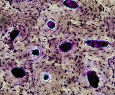
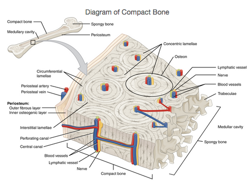
Bone tissue is distinctive and primarily extracellular materials. Compact and spongy bone are found in most bones; compact bone makes up the majority of the skeleton. The functional unit of compact bone is an osteon, which appear as adjacent tree rings. The cells (osteocytes), are surrounded by a calcified matrix of extracellular materials, but the tissue is living and served by blood vessels, lymph and nerves. Examine the slide and the model of bone tissue.
Hyaline Cartilage
Function: Support and protection, reduces friction
Locations: Nose, trachea and bronchi, ends of bones, ribcage, intervertebral disks
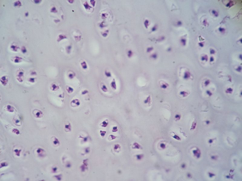
Hyaline cartilage has an extracellular gel-like matrix containing few/no visible fibers and scattered cells called chondrocytes, and overall appears less organized than bone. This tissue supports, protects, and minimizes friction where bones meet. Hyaline cartilage is found in the nose, trachea, ribcage, intervertebral disks and covers the ends of bones.
Skeletal muscle tissue
Function: Movement (voluntary)
Locations: Skeletal muscles
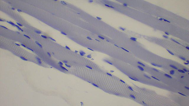
Skeletal muscle tissue contains cells filled with parallel fibers containing actin and myosin protein fibers. These fibers alternatively do/not overlap, causing the tissue to have a striated (striped) appearance like a candy cane. The nuclei are numerous (multi-nucleated), darker, and pushed to the margins of the cells. Identify the fibers, nuclei, and striations.
Gross Anatomy of Bones and the Skeletal system
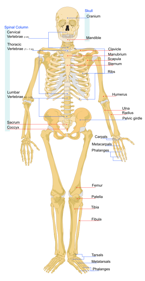
Bones (e.g. humerus) are organs since they contain >2 tissues including blood vessels, lymph vessels, and nerves, in addition to bone tissue. The human skeleton can be logically divided into the axial and appendicular divisions. The axial skeleton forms the main trunk, while the appendicular skeleton includes the bones of arms and legs (appendages), and the pectoral and pelvic girdles. In total, the adult skeleton usually contains 206 bones (80 axial and 126 appendicular). Among the 206 bones, there are 22 bones of the skull, 26 vertebrae, 12 pairs of ribs, and 30 bones in each appendage.
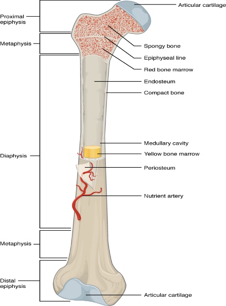
Observe a bone from a cadaver that has been cut longitudinally to reveal that it is actually hollow. Since this bone is dried out, it is still strong, but much less flexible than when alive. It contains compact bone along the sides and spongy bone at the ends. In life, the articular surfaces were covered with glossy articular cartilage (imagine the end of a chicken bone), the spongy bone was filled with red marrow and the medullary cavity was filled with yellow marrow (fat).
Some Major and Notable Bones / Bone Groups of the Body
Some basic bone descriptions
| maxilla: upper jaw | mandible: lower jaw | sternum: chest, ribs attach to it | clavicle: collarbone | scapula: shoulder |
| humerus: upper arm | radius: forearm (thumb side) | ulna: forearm | carpals: wrist | metacarpals: hand |
| phalanges: fingers | coxal bones: pelvis | sacrum: lower vertebrae (bones are fused) | coccyx: tailbone | femur: thigh |
| patella: knee | tibia: shin | fibula: lower leg (next to shin) | tarsals: ankle | metatarsals: foot |
| phalanges: toes |
* Are there more bones in the axial or appendicular division? ______________________
* Name 2 bones from the axial skeleton _____________ _________________
* Name 2 bones from the appendicular skeleton _____________ _________________
The following activity will help in learning the location and names of the bones. You will work as a group using the bones mixed up in tubs and reassemble them into (as complete as possible) a full skeleton. After assembling the skeleton, do not disassemble it until instructed to do so by you professor.
CSI Activity: a mass grave has been found and a mixture of bones recovered, but it is unclear how many victims there were. In order to determine the minimum number of victims, you must determine 1) how many bones of each type (e.g. femur) and 2) which side of the body they are from (Left or Right). Using the fully articulated skeletons at the front of the rooms as a guide, match each bone to the correct part of the body including L/R side. Use any articulation points, grooves, asymmetry, bone angles, and differences between the two ends of the bone to determine L/R. Recall that L/R is from the patient’s (or victim’s) perspective. Whichever bone(s) is the most abundant (L/R are counted separately, an exception is the vertebrae), represents the minimum number of victims. For example, if the most abundant bone is 4 right femurs, there are at least 4 victims. The first two vertebrae (atlas and axis, C1 and C2) are unique and may also be used to count victims.
* Complete the table. Before putting the bones back into the tubs, have your instructor verify your findings and review the bones you identified.
Crime Scene Investigation (CSI) Activity
A. Names of Bones
Ex.__femur_____________
1. ____________________
2. ____________________
3. ____________________
4. ____________________
5. ____________________
6. ____________________
7. ____________________
8. ____________________
9. ____________________
10. ___________________
11. ___________________
12. ___________________
13. ___________________
14. ___________________
15. ___________________
16. ___________________
17. ___________________
18. ___________________
19. ___________________
20. ___________________
B. # Left / # Right if known
Ex.____1 / 2_(at least 2 victims)
1. _________/________
2. _________/________
3. _________/________
4. _________/________
5. _________/________
6. _________/________
7. _________/________
8. _________/________
9. _________/________
10. _________/________
11. _________/________
12. _________/________
13. _________/________
14. _________/________
15. _________/________
16. _________/________
17. _________/________
18. _________/________
19. _________/________
20. _________/________
C. Minimum # of Bodies ____________
D. Explanation for determining minimum # (most numerous bone(s), which side of body): _______________________________________
E. Name the 5 regions of the backbone from top to bottom and list the number of vertebrae in each:
Skull – 1. 2. 3. 4. 5. – Pelvis
F. Number of vertebrae in each region (total of 26): , , , , _____
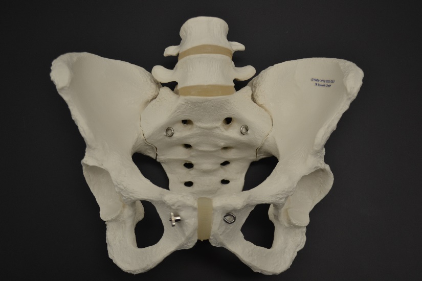
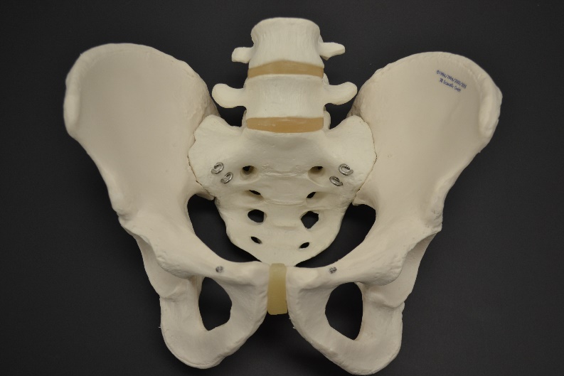
Observe the male and female pelvis on the cart or back counter, note any differences.
 ……… ……
__________ _________
* Label the pelvises above as male or female.
* What are two basic differences between M/F pelvis? 1.________________ 2. _______________
Muscular System
Skeletal muscles are under conscious (voluntary) control and appear striated (striped). Cardiac and smooth muscles are involuntary, and differ visually from skeletal muscle. Tendons are continuous with muscles and attach them to bones. The point of attachment that stays stationary when the muscle contracts is the origin, while the attachment that moves is the insertion. Since muscles shorten when they contract, they only pull. As a result, most muscles work in antagonistic pairs (particularly in the appendicular region)- when one contracts the other relaxes.
There are over 600 muscles in humans which are named based on their location, shape, size, movement, number of major parts or various other features. Examine the muscle model and study the images; learn the major “workout” and other muscles that your instructor indicates for the quiz.
Major and Notable Muscles of the Body
| Orbicularis oculi | External oblique | Flexor carpi group | Tibialis anterior |
| Orbicularis oris | Rectus abdominis | Extensor carpi group | Extensor digitorum longus |
| Masseter | Trapezius | Extensor digitorum | Gluteus maximus |
| Deltoid | Latissimus dorsi | Quadriceps femoris | Hamstring (Biceps Femoris) |
| Pectoralis major | Biceps brachii | Sartorius | Gastrocnemius |
| Serratus anterior | Triceps brachii | Adductor longus |
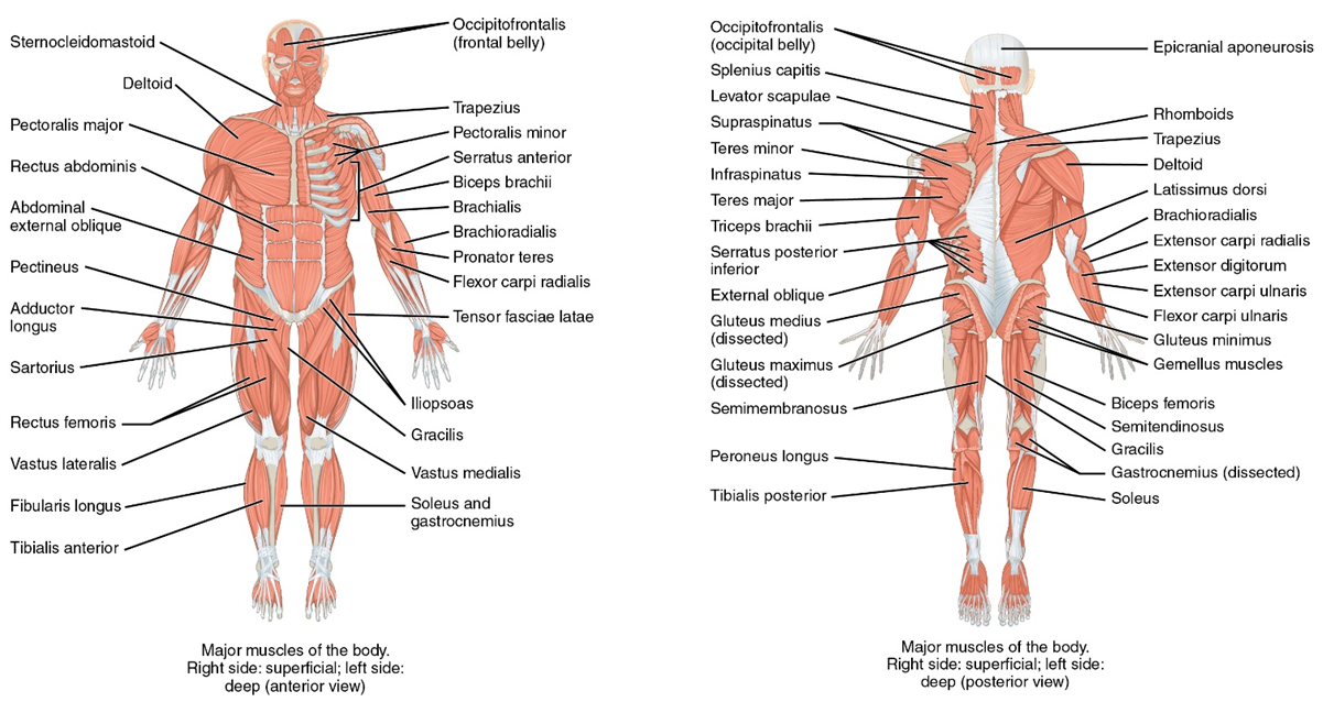
Some basic muscle descriptions
| o. oculi: circular muscle around eyes, blinking, winking | biceps brachii: upper arm muscle on front, bends arm at elbow |
| masseter: chewing muscle | triceps brachii: upper arm muscle on back, straightens arm at elbow |
| frontalis: forehead muscle raises brows | quadriceps femoris (quads): several muscles on front of leg, straightens leg at knee |
| deltoid: shoulder muscles, raises and lowers arms to front and side | sartorius: strap-like muscle running diagonally across thigh, moves thigh away from body |
| pectoralis major: chest muscles, bring arms forward | tibialis anterior: shin muscle, turns foot upward as when walking on heels |
| external oblique: muscles on your side running diagonally onto stomach | gluteus maximus: butt muscles, extends thigh back |
| rectus abdominis stomach muscles with fibers running vertically (6-pack abs), bends vertebral column | hamstrings (biceps femoris): several muscles back of leg, bends leg at knee |
| trapezius: sheet like muscle covering upper back, raises shoulders | gastrocnemius: calf muscles, turns foot downward |
| latissimus dorsi: (lats) beneath arms onto back, brings arms down and backward behind body |
Muscle movements
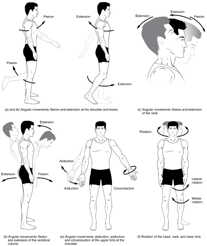
Muscle contractions are described based on their movement in relation to the joint or the midline of the body. The main types are flexion, extension, adduction, and abduction. Flexion: movement of jointed parts towards one another, Extension: movement of jointed parts away from each other, Adduction: movement of part toward body’s midline, Abduction: movement of part away from body’s midline.
* Answer the following:
The biceps brachii ________ the forearm.
The triceps brachii ________ the forearm.
The quadriceps femoris __________ the leg.
The biceps femoris _________ the leg.
The sartorius _________ the thigh.
The adductor longus ___________ the thigh.
Contraction Types
In isotonic contractions, the length of the muscle changes. In isometric contractions, the length of the muscle does not change.
To demonstrate an isotonic contraction, rest your left forearm on a table. Watch the anterior surface of your left upper arm while you slowly bend your elbow and bring your left forearm toward the upper arm. This is an isotonic contraction of the biceps brachii.
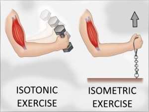
* What makes this contraction isotonic rather than isometric? __________________________________
To demonstrate an isometric contraction, place the palm of your left hand underneath a tabletop. Push up against the table while you have your right hand cupped over the anterior surface of your left upper arm so that you can feel the muscle there undergo and isometric contraction.
* What change did you notice in the firmness of the triceps brachii as it is contracted? ______________
* Did your hand or forearm move as you pushed up against the table? ___________________________
* Did this muscle’s fibers shorten as you pushed up against the table? __________________________
Contraction of muscle fibers
The striations in muscle fibers are due to locations of actin and myosin fibers. During contraction, actin filaments slide past and overlap more with myosin filaments. The result is shortening of the sarcomere, the functional unit of muscle. Examine the microscope structure of muscle fibers showing the shortening of the sarcomere.
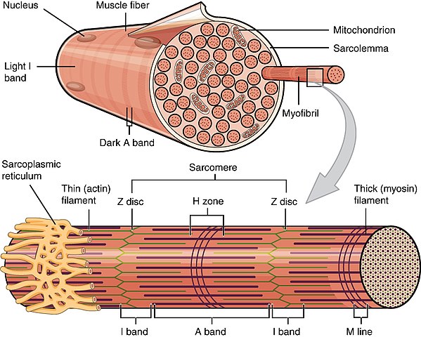
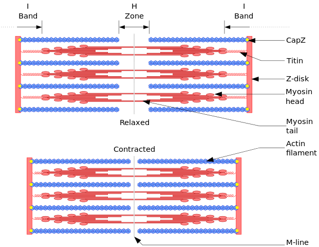
* The functional unit of muscle is called a(n) ______________________________________.
* What shortens during a muscle contraction: actin, myosin or the sarcomere? _____________________
Joints
The study of joints and articulations is arthrology, while studying the body’s movements is called kinesiology. Where bones meet, joints are generally immovable, slightly movable or freely movable (= synovial). There are six main types of synovial joints to know: 1. Ball and socket, 2. Condyloid, 3. Hinge, 4. Pivot, 5. Gliding, 6. Saddle. These joint types vary in their plane of movement, with ball and socket joints having three planes of movement. Joints are normally named by the articulations of the bones involved. For example, the elbow (a hinge) joint is called the humeroulnar joint.
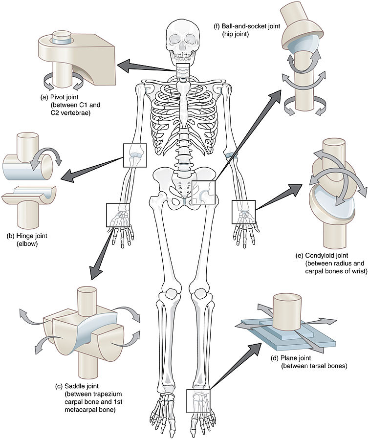
* Examine the models of the hip, shoulder, and knee joints. Note the following:
– Deep socket, stability of the hip joint– Shallower socket, greater range of movement of shoulder
– Overall instability of the knee– Ligaments serving the knee front to back & side to side
* Which ball and socket joint is more easily dislocated? __________________
Types of Joints in the Body
- Immovable – ex. sutures between skull bones
- Slightly Movable – ex. vertebrae and disks & coxal bones (pubic symphysis)
- Movable Joints (Synovial): name all bones involved in the joint
Complete the following using the word bank:
Terms: atlas, axis, carpals, coxal (=pelvis), femur (2), humerus (2), metacarpals (2), phalanges, radius (2), scapula, tibia (2), tarsals (2), ulna
1. Ball & Socket*
Shoulder (name 2 bones involved) _____________________…. Hip (2) __________________________
2. Condyloid*
Wrist (2) ______________________________…. Foot (2) ______________________________
Finger (2) ____________________________
3. Hinge*
Elbow (2) _________________________…. Knee (2) ______________________________
4. Pivot*
Lower arm (2) ___________________________…. Top two vertebrae (2) _______________________
5. Plane/Gliding Joint
Between wrist bones __________________Between ankle bones _________________
Image Credits:
Bone By https://commons.wikimedia.org/wiki/File:Compact_bone_-_ground_cross_section.jpg
Diagram of compact bone By OpenStax College – Anatomy & Physiology, Connexions Web site https://cnx.org/contents/FPtK1zmh@8.108:kwbeYj9S@4/Bone-Structure
Cartilage By https://commons.wikimedia.org/wiki/File:Cartilage01.JPG
Muscle tissues By 乌拉跨氪 – Own work, CC BY-SA 4.0, https://commons.wikimedia.org/w/index.php?curid=46401576
Long Bone By OpenStax College – Anatomy & Physiology, Connexions Web site https://cnx.org/contents/FPtK1zmh@8.108:kwbeYj9S@4/Bone-Structure
Human Skeleton Front By https://commons.wikimedia.org/wiki/File:1105_Anterior_and_Posterior_Views_of_Muscles.jpg
Male and Female Pelvises Photos By Jeffery Ray, author of this lab activity
Human muscles By OpenStax [CC BY 4.0 (https://creativecommons.org/licenses/by/4.0)], via Wikimedia Commons https://commons.wikimedia.org/wiki/File:1105_Anterior_and_Posterior_Views_of_Muscles.jpg
Muscle contractions by
Body Movements https://cnx.org/contents/FPtK1zmh@8.108:qCnsYyus@3/Types-of-Body-Movements
Isotonic/Isometric By http://isowalking.com/
Skeletal muscle fiber https://cnx.org/contents/FPtK1zmh@8.108:bfiqsxdB@3/Skeletal-Muscle
Sliding filament By Richfield, David (2014). “Medical gallery of David Richfield”. WikiJournal of Medicine 1 (2). DOI:10.15347/wjm/2014.009. ISSN 2002-4436. – Own work, CC BY-SA 3.0, https://commons.wikimedia.org/w/index.php?curid=2264027
Joints by By OpenStax College – Anatomy & Physiology, Connexions Web site. http://cnx.org/content/col11496/1.6/, Jun 19, 2013., CC BY 3.0, https://commons.wikimedia.org/w/index.php?curid=30131668
BI 102 Lab Worksheet: Musculoskeletal Name ___________________________ Section _______
1. How many bones in the adult human?_____ How many muscles in the adult human? ____
2. What are the two main divisions of the human skeleton?
1. _____________________________2.______________________________
3-4. Name the bone in the upper arm: General name for bones of the ankle:
Name the bone of the lower jaw: Name for collarbone:
5. Name & sketch the shape of any two types of vertebrate:
1. Type: 2. Type:
6. Name one location you would find a pivot joint:
7. Sketch the basic shape differences of
a female vs. a male pelvis (see front cart)→:
8-9. Give the everyday name for the following muscles:
Gastrocnemius: Deltoid: Masseter:
Gluteus maximus: Quadriceps femoris:
Trapezius:
10. Name & sketch the shape any two tissue types you observed under the microscopes (side/back counter):
1. Tissue: 2. Tissue:
