4 Chapter 4
LAB 4
Introduction to the Digestive System
Prepared by Jason R. Jones, University of North Alabama
OBJECTIVES
After completing these laboratory activities, you should understand / be able to:
- Recognize the organs of the digestive system, including those that are part of the alimentary canal or digestive tract, as well as those that are considered accessory organs, and their functions.
- The difference between mechanical digestion and chemical digestion.
- Define the terms catalyst, enzyme, and substrate.
- That enzyme activity can be affected by multiple factors, and examples of such factors affecting enzyme activity.
- Define the terms positive control and negative control, and know how/why they are important.
- Define the term peristalsis.
- Some of the major digestive enzymes involved in digestion of carbohydrates, proteins, and fats; know where these enzymes are produced, and the site of their actual function.
- Different factors that can affect the activity of digestive (and other) enzymes.
- The role of bile in the emulsification of fats.
INTRODUCTION
The digestive system is responsible for processing the food taken into the body and converting it to usable energy.
The process of digestion can actually be divided into two main types of digestion. The first of these is mechanical digestion, which is simply the breaking down of food into smaller pieces, but without any chemical changes to the food. This process is begun in the mouth, through the chewing action of our teeth. By chewing food into smaller pieces, this increases the surface area of the food. This larger surface area thus provides for faster and more efficient chemical digestion (which also begins in the mouth), in which various enzymes actually further break down the food chemically into smaller molecules that are more easily absorbed and used by our bodies for various functions. An enzyme is an organic molecule (usually a protein) that acts as a catalyst (a molecule that speeds up a particular reaction) for the breakdown of large molecules in our food into smaller “building block” molecules. Enzymes are usually very specific, and a given enzyme typically only speeds up a single type of reaction. Enzymes speed up reactions by binding to a substrate, which is one of the reactants in a chemical reaction.
In these exercises, you will familiarize yourself with the anatomy of the digestive system, including the organs of the alimentary canal (or digestive tract) and the various accessory digestive organs that also play important roles in digestion. You will also examine the activity of several digestive enzymes, as well as explore factors that may affect the activity of these enzymes.
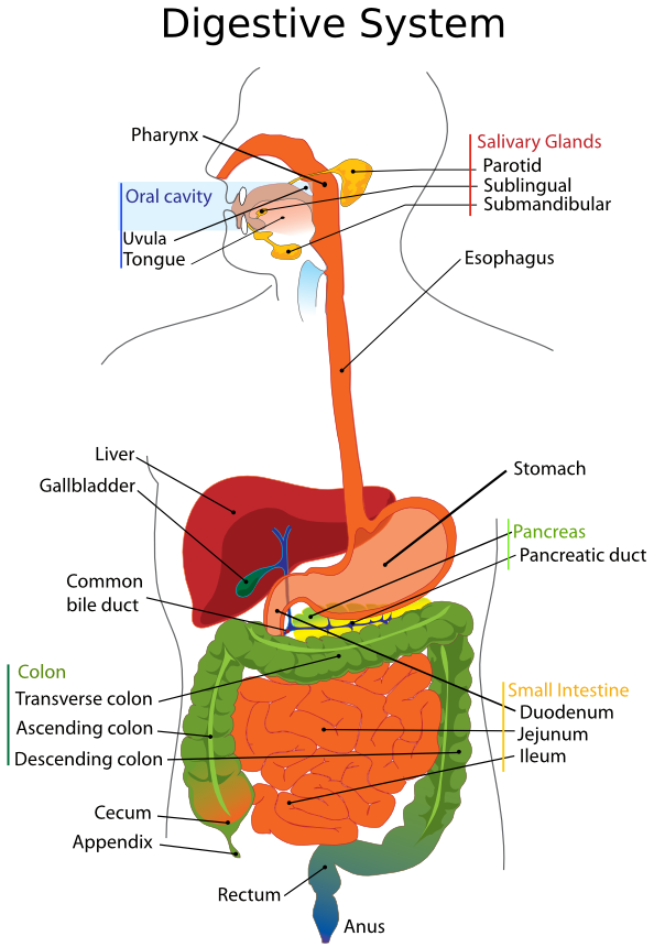
Figure 1. Organs of the digestive system
ACTIVITY I: Organs of the Alimentary Canal
When most people think of the digestive system, they typically think of the digestive tract, also known as the alimentary canal, though there are other organs that function in digestion (the accessory digestive organs, which we’ll discuss later). The alimentary canal consists of all of the organs of the digestive system through which food passes directly, and includes the oral cavity (mouth), pharynx (throat), esophagus, stomach, small intestine (subdivided into several regions), and large intestine (also subdivided into several regions). Each of these organs are hollow and can essentially be thought of as subdivisions of a single tube, with an opening at each end (mouth and anus). The overall structure of the organs of the alimentary canal is fairly similar, with each consisting of four basic layers. The inner surface of each of these organs is lined with a mucus membrane, consisting epithelial tissue and a thin layer of smooth muscle. This layer serves several functions, including secretion (mucus, enzymes, hormones, etc.), absorption, and protection from microbial invaders. Surrounding the mucus membrane layer is a layer of dense connective tissue containing blood vessels, lymph vessels, and nerves. The next layer of the alimentary canal (moving outward) is a double layer of smooth muscle tissue. This involuntary muscle tissue is responsible for movement of food along the digestive tract via a wave of contraction (called peristalsis) that moves sequentially along the length of the digestive tract. The outermost layer of the digestive tract consists of epithelial tissue and a thin layer of connective tissue. This outermost layer mainly serves to anchor the digestive organs in place, as well as reduce friction as the movable digestive organs slide against one another and along body cavity walls.
Food enters the alimentary canal through the oral cavity, or mouth. The oral cavity is bounded by the lips anteriorly (to the front), by the cheeks laterally (to the sides), and the hard palate (which has underlying bone) and soft palate (with no underlying bone) forming the roof of the mouth, and the tongue occupying the floor of the mouth. Inside the oral cavity are the gums, teeth, tongue, and openings to the salivary glands, which are accessory digestive organs. During chewing, the salivary glands release saliva, a watery mixture of mucus, cells, enzymes, and other molecules that moistens (and begins the process of digestion of) the chewed food, facilitating its compression into a mushy ball called a bolus. During swallowing, the tongue moves the bolus towards the back of the mouth towards the pharynx. (Swallowing is actually a fairly complex process, involving over 30 different muscles).
The pharynx (throat) is a passageway that contains openings leading into both the digestive tract (via the esophagus) and the respiratory tract (via the trachea). To prevent food from entering the respiratory tract, a flap of cartilage called the epiglottis folds down to close the glottis (opening of the trachea) during swallowing. To illustrate the activity of the epiglottis in its role of closing the respiratory tract during swallowing, follow the directions below.
- Place your index and middle fingers on your larynx (voice box). This is easier to see in males than in females, due to the enlarged laryngeal prominence (Adam’s apple), which is more pronounced in males. However, if you are a female, you should still be able to easily locate the large area of cartilage that marks the upper portion of the respiratory tract.
- With slight pressure against the larynx, swallow. You should be able to feel the larynx rise, as well as a slight posterior (backwards) motion, representing the epiglottis folding backwards to cover the opening of the trachea (windpipe).
- Answer the question on the worksheet at the end of this lab exercise.
After swallowing, the bolus of food then moves into the esophagus, which has no digestive or absorptive function, and simply serves as a passageway into the lower gastrointestinal tract. The esophagus contains smooth muscle, which contracts via wavelike peristaltic motion to move the bolus to the stomach. The opening between the esophagus and stomach is typically closed by a ring of involuntary smooth muscle called the gastroesophageal sphincter. Upon swallowing, when the wave of peristaltic contraction reaches this sphincter, it forces it open, allowing the bolus to enter the stomach.
To illustrate the activity of peristaltic contraction and the gastroesophageal sphincter, follow the instructions below.
- Take the provided stethoscope, and clean the earbuds with an alcohol swab.
- Have your partner use their fingers to locate their xiphoid process, which is the small, inferior-most (lower) bone of their sternum.
- Have your partner place the membrane of the stethoscope approximately one inch below this point.
- Use the provided small cup of water. While listening, have your partner take a fairly large mouthful of water, and swallow it.
- Listen closely, and you should hear two sounds. First, you should hear a splash, as the water strikes the closed esophageal sphincter. As soon as you hear this initial splash, start the provided timer.
- Continue to listen, and you should hear a second sound. As soon as the peristaltic wave of contraction of the esophagus reaches the sphincter, this should force it open, allowing the water to enter the stomach, which should produce a gurgling sound. When the gurgling sound is heard, stop the provided timer.
- Answer the questions in the worksheet at the end of this lab exercise.
The stomach is a J-shaped, organ that functions in both mechanical and chemical digestion. Look at the provided stomach model. The outer layer of the stomach consists of epithelial tissue, with a thick layer of smooth muscle just underneath. The muscle layer actually consists of three layers of smooth muscle: an outer layer of longitudinal muscle, a middle layer of circular muscle, and an inner layer of oblique smooth muscle (note that the names of each of these layers correspond to the direction in which the muscle fibers are arranged in those layers). The innermost layer of the stomach consists of simple columnar epithelial tissue arranged into ridges and folds, which also contain small pits. These ridges and folds assist with mechanical digestion of food, as contraction of the smooth muscle layers of the stomach churn food in the stomach, as well as pummel food against these ridges, further breaking the food into smaller pieces. These ridges/folds also increase the surface area of the stomach lining, allowing for the presence of a greater number of cells that produce various important molecules, such as mucus (which protects the lining of the stomach), hydrochloric acid (HCl, which is involved in chemical digestion), and digestive enzymes (discussed further later).
After further processing in the stomach, the mixture of partly digested food and gastric juices (acid, digestive enzymes, and mucus) passes through a valve called the pyloric sphincter into the small intestine, which is divided into three main regions (in order): duodenum, jejunum, & ileum. The first section directly connected to the stomach is the duodenum. The majority of the small intestine is the jejunum, and the terminal portion that connects to the large intestine is the ileum. The human small intestine in a cadaver, stretched out, would be ~20 feet in length. In life, muscle tone of the small intestine reduces its length to ~6 feet. The small intestine is the site of further digestion of food, as well as the site of nearly all absorption of nutrients into the bloodstream. Look at Figure 2 on the following page. Note that the small intestine is lined with numerous fingerlike projections called villi, which themselves are covered with even tinier fingerlike projections called microvilli. Both villi and microvilli serve to increase the surface area of the small intestine, creating additional surface area for absorption of nutrients.
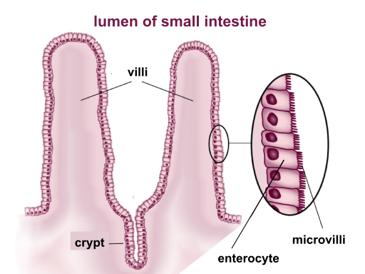
Figure 2. Microscopic view of the lining of the small intestine showing villi and microvilli. This work by BallenaBlanca (modified byMcortNGHH) is licensed under a Creative Commons Attribution 4.0 International (https://creativecommons.org/licenses/by-sa/4.0/deed.en).
The last segment of the small intestine, the ileum, empties into the large intestine (or colon). The large intestine plays very little role in the digestion of food, and primarily serves to reabsorb water from the digested food, as well as vitamin K produced by colon bacteria. Like the small intestine, is divided into several distinct regions. The first region of the large intestine, which is directly attached to the ileum of the small intestine, is a pouch shaped region called the cecum. At the posterior (lower) end of the cecum is a short, twisted, wormlike pouch called the appendix. Though it was previously thought to have little to no function, the appendix primarily serves as a reservoir for beneficial gut bacteria. Superior to (above) the cecum is the segment of the large intestine referred to as the ascending colon. The ascending colon then bends in an approximately 90 degree angle, after which the large intestine travels across the body as a region called the transverse colon, which also terminates in a near 90 degree angle, marking the beginning of the descending colon. At its lower end, the descending colon leads into an S-shaped curved portion of the large intestine called the sigmoid colon (“sigmoid” literally means “S-shaped”). The sigmoid colon leads into the vertical segment of the large intestine called the rectum, which serves as an area of storage of formed feces before its eventual elimination from the body through the anus, which has two circular sphincter muscles: an involuntary internal sphincter of smooth muscle, and a voluntary external sphincter of skeletal muscle. Answer the question about several regions of the large intestine on the worksheet at the end of this lab exercise.
Familiarize yourself with the location of each of the above regions of the digestive tract in Figure 1, as well as on the provided human torso models. On the worksheet at the end of this lab exercise, you will be asked to number the regions of the digestive tract in the order in which food moves through them throughout the digestion and excretion process.
ACTIVITY II: Structure & Function of Accessory Digestive Organs
Activity IIA: Mammalian Tooth Types and Mammal Dental Formulas
The process of mechanical digestion begins with the chewing action of the teeth breaking food into smaller pieces. Mammals (including ourselves) have different types of teeth adapted for different functions. Incisors, for example, are narrow-edged teeth towards the front of the mouth that are adapted for cutting. The canines are also teeth near the front of the mouth, and are typically pointed. The canines primarily are adapted for tearing food. In carnivores, the canines are generally very pronounced, but in omnivores and herbivores, they may be more similar in shape to the incisors. Moving towards the back of the mouth, the next type of tooth encountered are the premolars, also known as bicuspids (due to the presence of two cusps, or raised surfaces). Finally, the teeth farthest back in the mouth are known as the molars. Molars are distinguishable from premolars by a greater number of cusps on their surfaces. In omnivores and herbivores, both the premolars and molars are primarily adapted for crushing and grinding food, though in carnivores, they may be more modified for shearing meat.
The skull, and in particular, the teeth of a mammal can actually tell you a great deal about that organism, including the species from which the skull came, as well as some substantial insight into the dietary habits of that organism. Different species of mammals have different numbers and shapes of each type of tooth, and mammals are often described in terms of their dental formula. The dental formula of an animal is a way of displaying the number of each type of tooth found in both the upper (maxilla) and lower (mandible) jaw of its skull. To write the dental formula of a mammal, you count the number of teeth of each type (incisor, canine, premolar, and molar) on only one side of both the upper and lower jaw. The reason teeth are only counted on one side of each jaw is because mammals are bilaterally symmetrical, and should have the same number of each type of teeth on the other side of the jaw. Dental formulas are written in a format that looks somewhat like a fraction. Below is an example of how a dental formula is typically written:
ICPM/ICPM=U/L=T
However, in the dental formula of an actual mammal, the letters above the line would be replaced by the number of each type of tooth on one side of the upper jaw, and the letters below the line would be replaced by the number of each type of tooth on one side of the lower jaw (I = # of incisors, C = # of canines, P = # of premolars, and M = # of molars; U = total # of teeth in the upper jaw, L = total # of teeth in the lower jaw, and T = total # of teeth). To calculate the total number of teeth in the upper and lower jaws, you would add all the numbers above the line (for the upper jaw) or below the line (for the lower jaw), and multiply by two, because remember, dental formulas are written by counting the number of each type of tooth only one one side of the skull. For example, the dental formula of the Virginia opossum (Didelphis virginiana) would be written as follows:
5134/4134=26/24=50
This means that a Virginia opossum has a total of 26 teeth in its upper jaw (5 x 2 = 10 incisors; 1 x 2 = 2 canines; 3 x 2 = 6 premolars; 4 x 2 = 8 molars; 10 + 2 + 8 + 6 = 26), and a total of 24 teeth in its lower jaw (4 x 2 = 8 incisors; 1 x 2 = 2 canines; 3 x 2 = 6 premolars; 4 x 2 = 8 molars; 8 + 2 + 8 + 6 = 24), for a total of 50 teeth.
Using the provided model of an adult human skull, and Figure 3 below, see if you can determine the dental formula of an adult human, and write it in the appropriate space on the worksheet at the end of this lab exercise. Note that the provided model does not illustrate erupted wisdom teeth (3rd molars on each side on both the upper and lower jaws). In your determination of the human dental formula, include the wisdom teeth in your calculation.
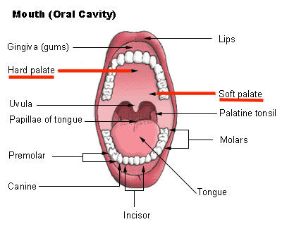
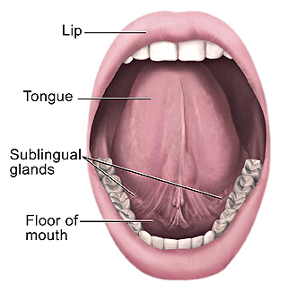
Figure 3. Anatomy of the oral cavity
Activity IIB: Location and Function of the Salivary Glands
There are three pairs of salivary glands that empty secretions into the oral cavity. The largest of these glands are the parotid glands, which are located in front of the ear, and which empty into the mouth just above the second molar. Another pair, the submandibular glands, which are located under the mandible, or lower jaw, have ducts that open on either side of the lingual frenulum, which is the membrane that attaches the tongue to the floor of the mouth. Another pair, the sublingual glands, is located underneath the tongue, and empty through several ducts towards the lateral (towards the side) aspects of the base of the tongue. Look at Figure 1, and note the location of each of these pairs of glands, and also see if you can locate any of them on the available torso models. Ask your partner to lift their tongue, and see if you can see the openings of the ducts of their submandibular and sublingual glands, and see if they can locate the openings of your ducts of these glands.
Though the amount of saliva that is produced by healthy individuals is debated, current estimates of saliva production range from about 0.75-1.5 liters of saliva per day, with the majority of saliva (70-75%) being secreted by the submandibular glands. The majority of the volume of saliva (about 99.5%) is water, but saliva also contains many other substances, such as mucus (consisting primarily of various proteins), that primarily serves the function of lubrication of the oral cavity and chewed food, as well as various enzymes, including some that function in digestion, as well as some that have antimicrobial activity.
One of the major digestive enzymes produced by the salivary glands is amylase, which is an enzyme that breaks complex carbohydrates (like starch) down into simple sugars. Amylase is also produced by the pancreas (and released into the duodenum; but this will be discussed later), but approximately 30% of starch digestion by amylase occurs in the mouth.
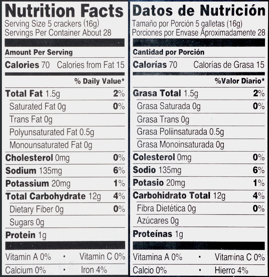
To illustrate the activity of salivary amylase, your instructor will provide you with a saltine cracker. Look at the nutritional label from a typical package of saltine crackers in Figure 3 to the right:
Notice that for a serving of crackers (16g), the majority of its mass is carbohydrates. However, notice the subcategories below the “Total Carbohydrate” information show a sugar content of 0g. This means the majority of saltine crackers consists of large complex carbohydrates, such as starch.
Now take the provided saltine cracker, and put it into your mouth, and begin chewing, but do not swallow. Continue chewing the cracker for at least 1 full minute, and notice the physical changes in the texture of the cracker, as well as any changes in flavor you may notice. Record this information in the worksheet at the end of this lab exercise.
Figure 4. Nutritional information for a typical package of saltine crackers.
Activity IIC: Additional Accessory Digestive Organs and their Functions
In addition to the teeth and salivary glands, there are several additional accessory digestive organs. Again, accessory digestive organs are organs through which food does not pass directly, but which contribute substantially to digestive function. These organs include the liver, gallbladder, and pancreas.
After the skin, the liver is the second largest organ of the body (weighing approximately 3 pounds), and also the body’s largest gland. The liver is located in the abdominal cavity, just below the diaphragm (the large sheet of muscle that separates the thoracic cavity, containing the heart and lungs, from the abdominal cavity), anterior to (in front of) the stomach, and towards the right side of the body. The liver performs many important functions, such as filtering blood from the digestive tract before returning it to the body’s general circulation, working with the pancreas to regulate blood glucose levels, synthesis of proteins important for blood clotting, and detoxifying chemicals and metabolizing medications. However, the primary digestive function of the liver is the production of bile. Bile is a mixture of cholesterol, bile salts, and a pigment called bilirubin, which is the result of the breakdown of hemoglobin. Small amounts of bilirubin are excreted in the urine, but the products of the breakdown of bilirubin are also responsible for the brown coloration of feces. The main function of bile is in the digestion of fat, but it is mostly involved in the mechanical breakdown, and not the chemical digestion of fat.
After being produced in the liver, bile is secreted into the gallbladder, a small green sac-like structure on the inferior (lower) surface of the liver, where it is stored until the ingestion of fats. When ingested food with substantial fat content enters the duodenum (the first section of the small intestine connected to the stomach), the gallbladder contracts, emptying bile into the duodenum. Again, bile does not chemically break down fat. Instead, bile helps emulsify fat with the gastric juices leaving the stomach and entering the duodenum. By emulsification, we refer to a more evenly distributed mixture of fluids that do not normally mix well. For example, imagine making a salad dressing of oil and vinegar. Normally, oil and vinegar do not mix well, since oil consists of nonpolar molecules, and vinegar (consisting of mostly water) consists of a polar solution. Because of the differences in polarity the molecules of water in the vinegar and the molecules of oil show no attraction to one another, causing them to separate into distinct layers, with the oil on top due to a lower density. However, in making such a salad dressing, one could add an emulsifying agent (such as a beaten egg or mustard), which contains molecules that have both polar and nonpolar regions. The polar regions of the emulsifying agent molecules are attracted to water molecules in the vinegar, and the nonpolar regions of the emulsifying agent molecules are attracted to the molecules of oil, and, after a good shake, this allows the oil molecules to be mixed evenly with the water molecules of the vinegar. This is exactly how bile allows emulsification of fats in the gastric juices, which are mostly water. The nonpolar regions of bile salt molecules essentially clump around tiny globules of oil molecules, with their polar regions facing outwards, and being attracted to water molecules in the gastric fluids. See Figure 5 on the following page for an example of this process.
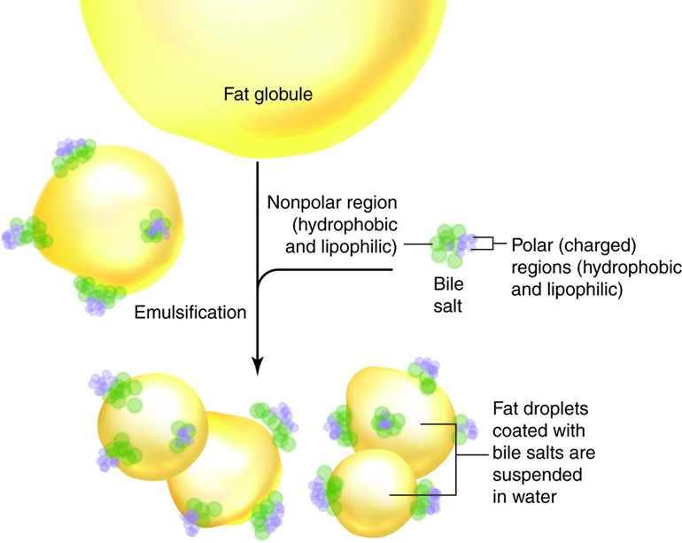
Figure 5. Emulsification of a fat globule by bile salts. This work by Cenveo is licensed under a Creative Commons Attribution 3.0 United States (http://creativecommons.org/licenses/by/3.0/us/).
Follow the directions below to illustrate the role of bile salts in the emulsification of fats.
- Obtain 2 test tubes.
- Label one tube “NB” to stand for “no bile”, and one tube “B” to stand for “bile”.
- To each tube, add 3 mL of distilled water.
- Next, add 3 mL of vegetable oil to each tube.
- To the tube labeled “B” only, add a small pinch of bile salts (available on the cart at the front of the lab).
- Now cover each tube with a small square of Parafilm, and cover the opening and Parafilm of each tube with your thumb.
- Shake each tube vigorously for at least 30 seconds, and return the test tubes to your test tube rack.
- After 10 minutes, observe each tube, and answer the questions on the worksheet at the end of this lab exercise.
An additional important accessory digestive organ is the pancreas, which is a small, triangular gland that is found between the spleen and the duodenum. The pancreas plays a role as both an endocrine gland (secreting hormones involved in blood sugar regulation directly into the bloodstream), but also as an exocrine digestive gland (secreting several important digestive enzymes, discussed later, into the duodenum). Additionally, as the highly acidic gastric juices from the stomach are passed into the duodenum, the pancreas also secretes sodium bicarbonate (NAOH), a highly basic fluid into the duodenum to neutralize the acid from the stomach.
Activity III: Digestive Enzymes and Factors Affecting their Function
Several digestive enzymes are produced by various organs of the digestive system, and are responsible for the chemical digestion of various organic macromolecules (chemically breaking the larger organic molecules into smaller building block molecules). Enzymes are organic molecules (typically proteins) that act as catalysts (molecules that speed up particular chemical reactions), without being used up in the process (enzyme molecules are not destroyed, and can be used again/recycled). The names of enzymes usually (but not always) end in the suffix “-ase,” so any time you see a biological reference to a molecule with that ending to its name, it is a safe bet that that molecule is an enzyme. Enzymes are usually very specific, and speed up only a single type of chemical reaction. Enzymes work by acting on a substrate (a molecule that temporarily binds to the enzyme molecule, with the enzyme speeding up a reaction in which that substrate is a reactant). Look at Table 1 below listing several major digestive enzymes, where they are produced, the site of their action, their substrates, and their products. Your instructor may ask you questions about some of these digestive enzymes on the quiz at the beginning of the following week’s lab.
Table 1. Several important digestive enzymes, and details about their production and activity.
|
Enzyme |
Site of production |
Site of action |
Substrate |
Products |
|
Amylase |
Salivary glands (salivary lipase) Pancreas (pancreatic lipase) |
Mouth Small intestine |
Starch & other polysaccharides |
Simple sugars |
|
Pepsin |
Stomach |
Stomach |
Proteins |
Large polypeptides |
|
Trypsin |
Pancreas |
Small intestine |
Large polypeptides |
Smaller polypeptides |
|
Lipase |
Salivary glands (salivary lipase) Pancreas (pancreatic lipase) |
Mouth Small intestine |
Triglycerides (fats) |
Monoglycerides and fatty acids |
In the following exercises, you will explore the activity of a few of these enzymes, as well as some factors that affect their function. If you remember the discussion of biochemistry in BI 101, you should be aware that the function of enzymes, as proteins, depends entirely on the shape of the enzyme (protein) molecule. Any factors (such as temperature, pH, etc.) that can change the shape of the enzyme (protein) molecule can change the function of that particular enzyme. Some changes in shape may make an enzyme more effective, some may make them less effective, and in some cases, some changes in shape may make the enzyme nonfunctional. When an enzyme’s shape has been changed in such a way that its function has been eliminated, we say that the enzyme has been denatured.
When conducting tests of an enzyme’s activity, it is important to test each treatment for the presence of the enzyme’s substrate, as well as the products of the reaction catalyzed by the enzyme. In addition, in using indicators to test for both substrate and product(s), it is also important to use both positive controls and negative controls. A positive control is a solution, which, before testing, we know DOES contain the molecule of interest. A negative control is a solution, which, before testing, we know DOES NOT contain the molecule of interest.
Activity IIIA: Effects of Time and Temperature on Starch Hydrolysis (breakdown) by Amylase
Amylase is an enzyme (produced by both salivary glands and the pancreas) that digests starch and other complex carbohydrates into simple sugars. Amylase breaks bonds between sugar molecules via hydrolysis, a reaction with water. In this activity, you will explore the roles that both time and temperature play in the activity of amylase with (starch) and its products (simple sugars).
To conduct a test for the presence of starch, you will add 4 drops of Lugol’s iodine (IKI) to the tubes as instructed below. A positive test for starch will result in the development of a blue-black color, while a negative test for starch will result in an amber color.
To conduct a test for the presence of simple sugars, you will add a dropper full of Benedict’s solution to the tubes as instructed below, and then place the tubes you are testing for sugars in a boiling water bath for 2 minutes. A positive test for sugars will result in a color change indicating the relative amount of sugar in the sample as follows: green (small amount of sugars), orange/yellow (moderate amount of sugars), or red (large amount of sugars). A negative test for sugars will result in the maintenance of the blue coloration of the Benedict’s solution after boiling.
- For this activity, you will need to label a total of 15 test tubes. Label one tube “BA”, and the other 14 tubes A1-A14.
- Prepare a boiling water bath in a 400 mL beaker on your hot plate.
- To the tube labeled “BA”, add 5 mL of the provided amylase solution.
- Place the tube labeled “BA” in a boiling water bath in a 400 mL beaker for 15 minutes.
- After boiling the tube labeled “BA”, remove it from the boiling water bath, and return it to your test tube rack to cool.
- After the “BA” tube has cooled, add the appropriate amounts of the appropriate solutions listed in the table on the following page, and conduct the appropriate tests (for starch or sugars) at the time specified in the table for each tube, and record your results for each test (positive or negative) in Table 2 below.
Table 2. Data from exercise on the effects of time and temperature on amylase activity.
|
Tube # |
Solutions to add |
Test to Conduct |
Time |
Results (+ or -) |
|
A1 |
2 mL distilled H2O |
Starch |
Immediately |
|
|
A2 |
2 mL distilled H2O |
Sugar |
Immediately |
|
|
A3 |
2 mL starch solution |
Starch |
Immediately |
|
|
A4 |
2 mL starch solution |
Sugar |
Immediately |
|
|
A5 |
2 mL glucose solution |
Starch |
Immediately |
|
|
A6 |
2 mL glucose solution |
Sugar |
Immediately |
|
|
A7 |
1 mL starch + 1 mL amylase |
Starch |
Immediately |
|
|
A8 |
1 mL starch + 1 mL amylase |
Sugar |
Immediately |
|
|
A9 |
1 mL starch + 1 mL amylase |
Starch |
After 30 min |
|
|
A10 |
1 mL starch + 1 mL amylase |
Sugar |
After 30 min |
|
|
A11 |
1 mL starch + 1 mL boiled amylase (BA) |
Starch |
Immediately |
|
|
A12 |
1 mL starch + 1 mL boiled amylase (BA) |
Sugar |
Immediately |
|
|
A13 |
1 mL starch + 1 mL boiled amylase (BA) |
Starch |
After 30 min |
|
|
A14 |
1 mL starch + 1 mL boiled amylase (BA) |
Sugar |
After 30 min |
|
Using your results from the experimental activity above, answer the questions on the worksheet at the end of this lab exercise.
BI 102 Lab Worksheet: Digestion Name _________________________________ Section _______
ACTIVITY I: Organs of the Alimentary Canal
1. Why is the closure of the trachea by the epiglottis an important event that occurs during swallowing?
2. The duration between the sounds heard after my partner swallowed a mouthful of water was ______ seconds.
3. Given that the esophagus is approximately 25 cm in length, it can be estimated that the peristaltic wave of muscular contractions of the smooth muscle of the esophagus moves at a velocity of ______ cm/s.
4. Look at Figure 1, as well as the provided human torso model. After tracing the path of food through the digestive tract, describe how the ascending colon, transverse colon, and descending colon got their names.
5. Number the regions of the digestive system listed below in the order in which food moves through them, with “1” being the first region which food passes through:
_____ Anus
_____ Ascending colon
_____ Cecum
_____ Descending colon
_____ Duodenum
_____ Esophagus
_____ Ileum
_____Jejunum
_____ Oral cavity
_____ Pharynx
_____ Rectum
_____ Stomach
_____ Transverse colon
Activity IIA: Mammalian Tooth Types and Mammal Dental Formulas
6. Write the dental formula of an adult human below.
___________________ = _____ = _____
7. Describe the changes in texture and flavor you experienced after chewing the saltine cracker, and attribute these changes to specific components found in saliva.
Activity IIC: Additional Accessory Digestive Organs and their Functions
8. Describe the appearance of the contents of the tube with oil and water only (“NB”), and the tube with oil, water, and bile salts (“B”). How do the differences in the appearances of the tubes’ contents reflect the activity of bile?
Activity IIIA: Effects of Time and Temperature on Starch Hydrolysis by Amylase
9. What is the significance of tubes #A1 & #A5?
10. What is the significance of tubes #A2 & #A4?
11. What is the significance of tube #A3?
12. What is the significance of tube #A6?
13. Compare your results for your tests of tubes #A7-A10. What does this tell you about the role that time plays on enzyme activity?
14. Compare your results for your tests of tubes #A11-A14. What does this tell you about the effect of extreme temperature (boiling) on the activity of amylase?
