6 Chapter 6
LAB 6
Introduction to the Nervous System
Prepared by Jason R. Jones, University of North Alabama
OBJECTIVES
After completing these laboratory activities, you should understand / be able to:
- Define the terms central nervous system, peripheral nervous system, neuron, neuroglia, synapse, neurotransmitter, nerve, effector, gray matter, white matter, spinal reflex.
- Identify and label the following parts of a neuron, as well as know their functions: cell body, dendrite, axon, axon terminal, Schwann cell, myelin sheath.
- Explain the different functions of the following: sensory neuron, interneuron, motor neuron.
- List the parts found in each of these three major regions of the brain, and their functions: forebrain, midbrain, hindbrain
- Locate and identify the following structures in the brain, as well as know their functions:
- cerebrum, ventricles, thalamus, hypothalamus, pituitary gland, cerebellum, pons, medulla oblongata
- The following lobes of the cerebrum, and be able to list their major functional roles: frontal lobe, parietal lobe, occipital lobe, temporal lobe,
- The basics of how spinal reflexes work, and be able to give an example of a spinal reflex observed in lab.
INTRODUCTION
The vertebrate nervous system is comprised of the brain, spinal cord, and the body’s network of nerves. However, the parts of the nervous system are often grouped into two main divisions. The central nervous system (CNS) consists of the brain and spinal cord, while the cranial nerves, spinal nerves make up the other division, the peripheral nervous system (PNS). Although most organs are made up of almost all four major tissue types (epithelial, connective, muscle, and nervous), the functional parts of the organs of the nervous system consist of nervous tissue (though there are some associated connective tissues). The primary function of the nervous system is the rapid transmission of signals throughout the body, allowing quick detection of and responses to changes in an organism’s internal and/or external environment.
In these exercises, you will familiarize yourself with the anatomy of the nervous system, starting at the cellular and tissue level. You will continue this exploration into higher levels of organization, and examine the anatomy of nerves, the brain, and spinal cord. You will also investigate the phenomenon of spinal reflexes, and learn how spinal reflexes result in physical responses without initial involvement of or processing by the brain. Finally, you will measure your average reaction time, and may be asked to compare it to others in your lab section. Your instructor may wish to compare averages of male & female reaction times, and/or turn the reaction time activity into a contest of sorts.
ACTIVITY I: Cells of Nervous Tissue
Nervous tissue consists of two main types of cells: neurons and neuroglia (aka glia or glial cells). With regards to the function of transmission of signals throughout the body, that role is performed by the neurons, which are specialized cells that transmit electrical and chemical signals. There are three main regions of a typical neuron. The part of the neuron that contains the nucleus is called the cell body. Extending from the cell body may be one or several projections. Neurons can be classified based on the number of projections extending from the cell body. Unipolar neurons have a single projection (which divides into two branches) extending from the cell body. Bipolar neurons have two projections: a branch-like dendron, which carries signals to the cell body, and a long axon, which carries signals away from the cell body. Most neurons, however, are multipolar neurons, with multiple branch-like dendrites that carry messages to the cell body, and a single axon that carries signals away from the cell body. See Figure 1 for examples of unipolar, bipolar, and multipolar neurons. Also see Figure 2 for a more detailed look at the anatomy of a multipolar neuron.
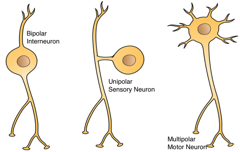
Figure 1. Examples of bipolar, unipolar, and multipolar neurons. Source: http://open.umich.edu/education/med/resources/second-look-series/materials
Notice in Figure 1, the axons of each type of neuron have button-like expanded regions at their tips. These regions are referred to as the axon terminals or terminal buttons. These terminals form junctions, called synapses, with other cells. Neurons can be classified based on the basis of the direction in which they are transmitting signals. Sensory neurons are neurons that detect stimuli (such as pain, heat, light, chemicals, etc.), and convert this information into electrical signals that are carried towards the central nervous system for processing. Motor neurons are neurons that carry processed signals in a direction from the central nervous system to an effector (such as a muscle or gland, which acts in response to this processed signal). However, there are other types of neurons, called interneurons or relay neurons, which carry information between sensory and motor neurons. See Figure 3 for examples of a sensory neuron, an interneuron, and a motor neuron. When an electrical signal (called an action potential) reaches the end of an axon, this causes the release of neurotransmitters, which are molecules that affect the activity of the other cells that meet with the neuron at the synapse. See the expanded view of a synapse in Figure 2, illustrating the release of neurotransmitter molecules from an axon terminal, and binding of the neurotransmitter to receptor proteins on the surface of a cell after the synapse. Most neurons form synapses with other neurons, and the neurotransmitters released from the pre-synaptic neuron (the neuron before the synapse) at those synapses can either make the post-synaptic neuron more likely to fire an action potential (excitatory) or less likely to fire an action potential (inhibitory). However, synapses also occur between neurons and muscle fibers, neurons and glands, and neurons and blood vessels. Neurotransmitters released from synapses between a neuron and a muscle fiber result in contraction of the muscle fiber involved. Neurotransmitters released from synapses between a neuron and a gland result in secretion of substances from the involved gland. Some neurotransmitters, however, are secreted directly from axon terminals into the bloodstream. See Figure 4 for some examples of several types of synapses between neurons and other cells.
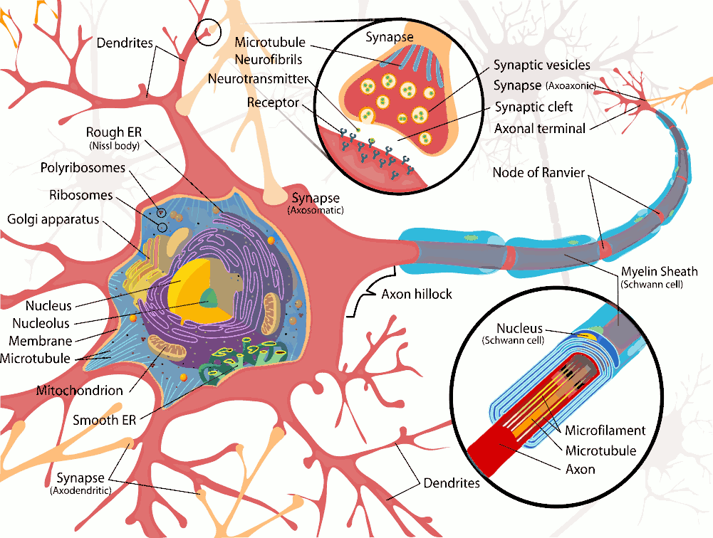
Figure 2. Anatomy of a typical multipolar neuron.
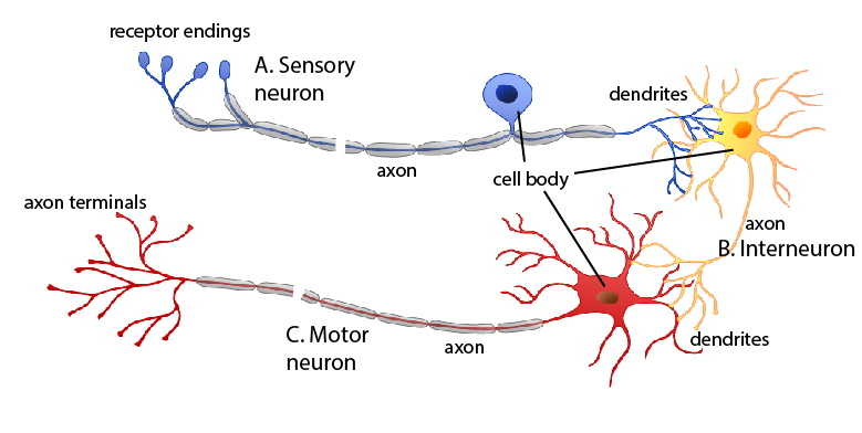
Figure 3. Illustration of a sensory neuron (A), an interneuron (B), and a motor neuron (C). Source: OpenStax at https://cnx.org/contents/pMqJxKsZ@6/Nervous-System

Figure 4. Examples of several types of synapses. Source: Blausen.com staff (2014). “Medical gallery of Blausen Medical 2014“. WikiJournal of Medicine 1 (2). DOI:10.15347/wjm/2014.010. ISSN 2002-4436
Though neurons are the cells that play the important role of signal transmission, several other important functions are performed by neuroglia, also known as glia or glial cells. Though they are not involved in signal transmission like neurons, glia have several functions that provide support to neurons in various ways. One function of glia is to act as the “glue” of nervous tissue, surrounding neurons and holding them into place. Glia also provide oxygen and nutrients to neurons, helping to keep them alive. Some glial cells also insulate neurons from other neurons by coating them with their cell membranes. Finally, some glial cells are capable of phagocytosis (“cell eating”), and can remove pathogens and dead neurons from nervous tissue. See Figure 5 for examples of several types of glial cells found in both the central and peripheral nervous system.
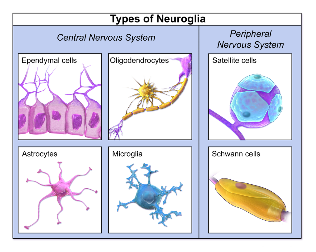
Figure 5. Types of neuroglia. Source: 1
You will not have to learn all of the different types of glial cells, but there is one type found in the peripheral nervous system (nerves outside of the brain and spinal cord that carry signals from the central nervous system to muscles, glands, and organs) that you should know. These types of cells are called Schwann cells, which can be seen in the bottom right of Figure 5, but also wrapped around the axon of the multipolar neuron shown in Figure 2. Schwann cells cover the surface of neurons in the PNS with a fatty substance known as myelin, providing a coating to the axon known as the myelin sheath. The function of the myelin sheath is to provide insulation to the axon of PNS neurons. Think about the myelin sheath as being analogous to a rubber coating on electrical wires. However, the myelin sheath does not cover the entire axon. There are gaps between the Schwann cells surrounding the axon where there is no myelin. These gaps are known as the nodes of Ranvier. These gaps in the insulating myelin facilitate more efficient transmission of electrical signals (action potentials) along the axon. Since these regions are not insulated, that allows the electrical signals passed along the axon to jump from node (non-insulated region) to node, instead of having to travel along the length of the axon continuously. This allows signals to be carried along PNS neurons much more quickly.
*Examine the neuron model, and answer the questions on the worksheet at the end of this lab exercise.*
Next, *Place the microscope slide of nervous tissue on the stage of a compound light microscope*
Starting with the scanning power (4x) objective, get the tissue on the slide into as sharp focus as possible using the coarse focus knob. After getting the tissue in focus on the scanning power objective, move up to the low power (10x) objective, and adjust the focus (if necessary) using the fine focus knob. Finally, move up to the high-power (40x) objective (providing a total magnification factor of 400x), and re-adjust fine focus as needed. You will likely see plenty of glia on the slide, but try to locate a neuron in this tissue. This may require scanning around the slide using the stage adjustment knobs. You may also need to make adjustments to the light intensity using the rheostat on the microscope, as well as to the condenser to best visualize the cells in this tissue. Once you have located a clearly visible neuron, go to the worksheet at the end of this lab exercise, and answer the appropriate questions regarding the micrographic view of this nervous tissue.
ACTIVITY II: Anatomy of the Brain
The brain is an organ seen in all vertebrates, and many invertebrates. Vertebrate brains vary among different vertebrate groups, but overall share the same basic structures. The brain is the enlarged anterior portion of the spinal cord, and with the spinal cord, as the central nervous system, acts as the main control center for the rest of the body.
The brain can be divided into three main regions: the forebrain, midbrain, and hindbrain, each of which consist of several other subregions/divisions. Using Figures 6-12 on pages 6-9 and Table 1 on the following page, you should familiarize yourself with several regions of the brain and their functions, and be able to identify them on the provided brain models.
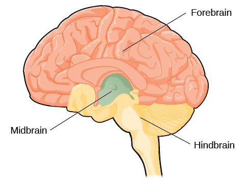
Figure 6. Major divisions of the brain. Source: OpenStax at https://archive.cnx.org/contents/fc8a38cc-fd1c-44cc-b91d-726fcfa62165@7/the-brain-and-spinal-cord
Table 1. Parts of the brain and their functions.
|
Brain Part |
Major Functions |
|
Forebrain Cerebrum Frontal lobe Central sulcus Parietal lobe Temporal lobe Olfactory bulb Occipital lobe Corpus callosum Thalamus Hypothalamus |
Higher order thinking and sensory processing Thought, problem solving, voluntary muscle control speech production Separates frontal lobe from parietal lobe Processing sensory input, sensory discrimination, body orientation Sound reception, expressed behavior, speech comprehension, memory Perception of smell Visual reception and interpretation Connects L/R hemispheres; allows communication between L/R brain Relays sensory info to cerebrum; regulation of sleep/consciousness Homeostasis (blood pressure, body temp, etc); pain/pleasure centers
|
|
Midbrain |
Responses to visual stimuli; motor coordination; eye movement |
|
Hindbrain Cerebellum Pons Medulla oblongata |
Essential body functions Equilibrium/balance & motor coordination Relays messages between cerebrum & cerebellum Control of heart, blood pressure, breathing; coughing, vomiting, sneezing, and swallowing reflexes |
|
Other Brain Parts to Know Ventricles Pituitary gland |
Filled with cerebrospinal fluid (acts as a shock absorber for CNS; provides nutrients to brain & spinal cord, and removes wastes from CNS) “Master gland” that produces hormones that control other glands of the endocrine system (thyroid, adrenal glands, gonads, etc.) |
Note that the brain stem is not mentioned in the table above. However, the brain stem, which controls the most basic body functions, consists of three main regions mentioned in Table 1: the midbrain, pons, and medulla oblongata.
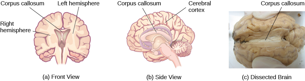
Figure 7. Illustration of the left and right cerebral hemispheres, and the corpus callosum, which connects them. Source: OpenStax at https://archive.cnx.org/contents/fc8a38cc-fd1c-44cc-b91d-726fcfa62165@7/the-brain-and-spinal-cord
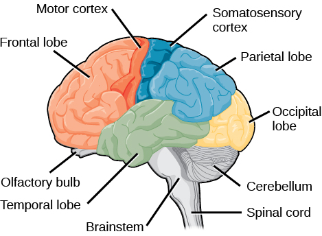
Figure 8. Lobes and cortices of the cerebrum. Source: OpenStax at https://archive.cnx.org/contents/2337db2c-8336-4955-bfd8-f57d3b9deaa4@2/human-biology-chapter-17-4-the-central-and-peripheral-nervous-systems
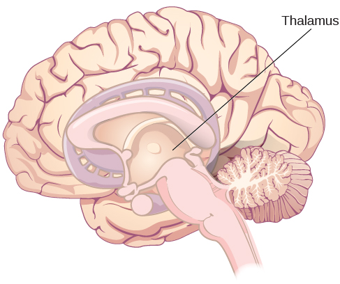
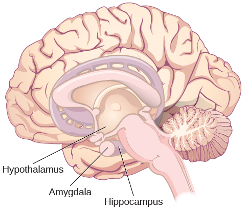
Figure 9. Other regions of the forebrain. Source: OpenStax at https://archive.cnx.org/contents/fc8a38cc-fd1c-44cc-b91d-726fcfa62165@7/the-brain-and-spinal-cord
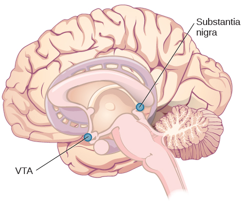
Figure 10. Structures of the midbrain. Source: OpenStax at https://archive.cnx.org/contents/fc8a38cc-fd1c-44cc-b91d-726fcfa62165@7/the-brain-and-spinal-cord
You will not need to know the structures illustrated in Figure 10. However, you should be aware of the basic location and structure of the midbrain. The midbrain is located deep within the brain, between the forebrain and hindbrain. The midbrain is primarily involved in movement, including eye movement, the processing of visual and auditory information, sleeping/waking cycles, and alertness/arousal. Additionally, the two regions illustrated in Figure 10, the ventral tegmental area (VTA) and the substantia nigra, contain cells that produce the neurotransmitter dopamine, which is involved in regulation of mood, including the perception of “rewarding” stimuli. Dopamine is also implicated in addiction, as release of dopamine occurs upon engaging in behavior in search of stimuli perceived as rewarding. Additionally, degeneration of these regions of the brain is also associated with progression of Parkinson’s disease.
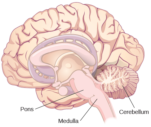
Figure 11. Structures of the hindbrain. Source: OpenStax at https://archive.cnx.org/contents/fc8a38cc-fd1c-44cc-b91d-726fcfa62165@7/the-brain-and-spinal-cord
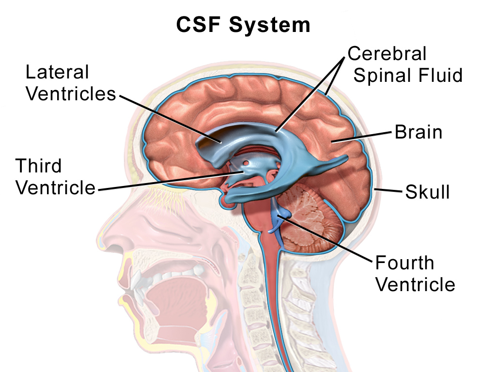
Figure 12. Illustration of the ventricles of the brain (hollow chambers containing cerebrospinal fluid, which acts as a shock absorber for the CNS, and provides nutrients to and removes wastes from the brain and spinal cord). Source: 1
Using the provided brain model, Figures 6-12, and Table 1, answer the questions on the worksheet at the end of this lab exercise.
ACTIVITY III: Anatomy of the Spinal Cord
In addition to the brain, the spinal cord is also part of the central nervous system. It is continuous with the medulla oblongata of the brain stem, and exits the base of the skull through a large hole called the foramen magnum. The spinal cord then passes through central holes in the vertebrae called the vertebral foramina, and extends down to the level of the first or second lumbar vertebra. Though not shown in Figure 13 below, the spinal cord is hollow, with a central canal filled with cerebrospinal fluid (CSF).
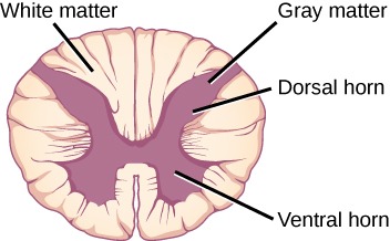
Figure 13. Cross section through the spinal cord showing gray matter (containing cell bodies and interneurons) and white matter (containing axons). Source: OpenStax at http://tea.cnx.org/contents/Rf2UxDVq@4/The-Central-Nervous-System
Using Figure 13 and the information in this exercise, answer the questions on the worksheet.
ACTIVITY IV: Anatomy of Nerves
The peripheral nervous system (PNS) consists of the cranial nerves (of which there are twelve pairs; illustrated on the provided brain models with Roman numerals), and the spinal nerves, which are also paired. Look at the provided model of the vertebral column, and count the number of roots of the spinal nerves (on the model, yellow structures sticking out laterally between vertebrae). Note that these models do not show the last sacral nerve (S5) or the coccygeal nerve (C0). Answer the question on spinal nerves on the worksheet. Remember to add the two pairs of nerves not shown on the model!
Nerves consist of bundles of axons of neurons grouped together in bundles called fascicles, each of which is surrounded by a layer of tissue called the perineurium. The fascicles are also held together in larger clusters of bundles by a layer of dense connective tissue (the epineurium) to make up the entire nerve. Look at Figure 14. Note that nerves are also vascularized (supplied by blood vessels), which serves to deliver oxygen and nutrients to neurons in the nerve, and to remove carbon dioxide and other wastes from those cells. After examining Figure 14 closely, look at the (somewhat crude, but rather effective/illustrative model of nerve anatomy on the cart at the front of the room. Then answer the questions on the worksheet at the end of this lab exercise.
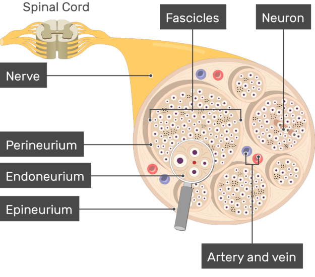
Figure 14. Illustration of the basic anatomy of a nerve. Source: https://www.getbodysmart.com/nerves/nerve-structure
ACTIVITY V: Spinal Reflexes
Reflexes are predictable, involuntary, and rapid responses to various environmental stimuli without said stimuli being first processed by the brain. Reflexes involve transmission of signals between two to three types of neurons, which were already discussed in Activity I: sensory neurons, interneurons (in some cases), and motor neurons. When an external stimulus (like stepping on a sharp nail) stimulates a sensory neuron, this information is relayed to the central nervous system (in some cases, to the brain stem, but in many cases, to the spinal cord). The signal from the sensory neuron is then relayed to an interneuron, and then to a motor neuron, or in some cases, directly from the sensory neuron to a motor neuron with no interneuron in between. This motor neuron then transmits the signal to an effector (such as a muscle), triggering the response. During this process, the response occurs before the stimulus is actually detected and processed by the brain, as the signal reaches the effector before the information is sent from the spinal cord to the brain. Figure 15 below illustrates the basic pathway of signal transmission in a spinal reflex arc.
In this exercise, you will explore a reflex with which you may already be familiar. If your doctor has ever tapped your knee with a rubber hammer, he or she did so to check transmission of signals between sensory neurons, the spinal cord, and motor neurons. This particular reflex is called the patellar reflex, or the knee jerk reflex. When your knee is struck with a reflex hammer, this causes a tendon, which is attached to both your patella (kneecap) and the quadriceps femoris muscle, to be stretched. This stretching stimulates receptors to generate electrical impulses that are passed from sensory neurons to the spinal cord, and then to motor neurons that stimulate the quadriceps femoris muscle itself. Figure 16 on the following page illustrates the pathway of signal transmission in the patellar spinal reflex arc.
To illustrate the patellar reflex, sit on the edge of your lab bench, so that your legs hang freely. Your partner will then take a rubber reflex hammer, and strike the lower edge of your kneecap with the pointed side of the hammer. If you do not initially exhibit a response to the strike of the hammer, your partner should try striking around the patella in slightly different locations until a response is observed. After observation of your response, switch places with your partner, and see if you can elicit a reflex response from them, as well.
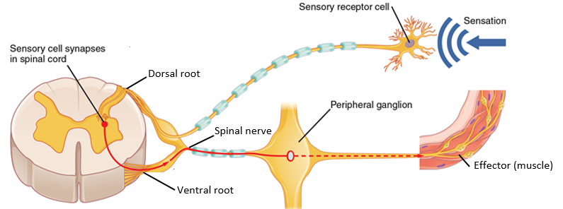
Figure 15. Illustration of signal transmission in a spinal reflex arc. Note that the sensory information is carried to the spinal cord through the dorsal root (on the back/upper side of the body), and relayed through interneurons (in some, but not all occasions) in the gray matter (not shown, but represented by the red dot in this figure), and back to the effector (a muscle) via the ventral root (on the front/lower side of the body) through a spinal nerve. Modified from OpenStax at https://archive.cnx.org/contents/a4ca89c4-ab19-492f-a910-8f0f7867999f@6/nervous-system
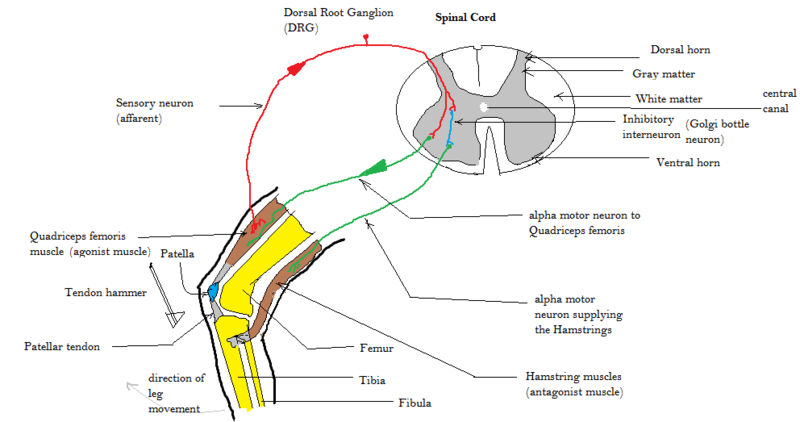
Figure 16. Illustration of the reflex arc of the patellar (knee jerk reflex). Source: Amiya Sarkar, from https://commons.wikimedia.org/wiki/File:Patellar_tendon_reflex_arc.png, licensed under Creative Commons Attribution-Share Alike 4.0 International license.
ACTIVITY VI: Measurement of Reaction Time
Follow the directions below for determination of you and your partner’s average reaction times:
- Your partner will stand in front of you while you are seated, and will hold the provided reaction time stick in the “Release” area between their thumb and forefinger.
- You should hold your thumb and forefinger of your dominant hand open about an inch apart and on each side of the “thumb line” on the reaction time stick, with your thumb closest to you, and your forefinger on the side of the reaction time stick farther away from you.
- Let your partner know when you are ready. Then, at some unspecified point, your partner will release the reaction time stick.
- You should attempt to catch the reaction time stick between your thumb and forefinger as quickly as possible.
- You can read your reaction time in milliseconds using the numbered markings on the reaction time stick.
- Record this reaction time in the space marked “RT1” on the worksheet at the end of this lab exercise.
- You should repeat this procedure for a total of 10 reaction time measurements, recording each reaction time on the worksheet.
- After your 10 trials, you and your partner will switch places, with you holding the reaction time stick, and your partner attempting to catch it as quickly as possible.
- Repeat for a total of 10 reaction time measurements for your partner, as well.
BI 102 Lab Worksheet: Nervous I Name _________________________________ Section _______
ACTIVITY I: Cells of Nervous Tissue
- ______________________also known as nerve cells, are the functional units of the nervous system.
- Using the model of a multipolar neuron, write the number from the model corresponding to the appropriate structures in the space below:
_____ Axon
_____ Dendrites
_____ Linked Schwann cells at node of Ranvier
_____ Nucleus
_____ Schwann cell (with nucleus)
_____ Synaptic terminal (axon terminal)
ACTIVITY II: Anatomy of the Brain
3. The most basic functions of the body like heartbeat and breathing are controlled by which part of the brain that includes the medulla and the pons? (damage/swelling in this part of the brain is often fatal)
4. Using the figure, match the letters to the correct cerebral lobes below, as well as their functions:
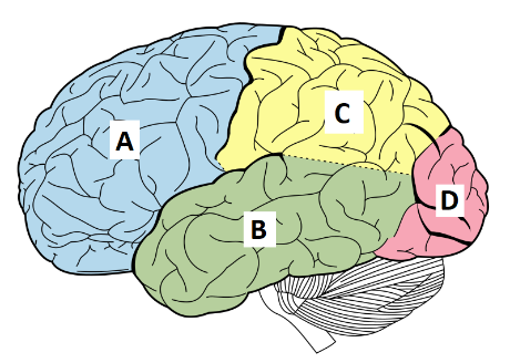
_____ Frontal lobe
_____ Occipital lobe
_____ Parietal lobe
_____ Temporal lobe
_____ Movement, thought, language production
_____ Auditory reception, behavior, expressed speech, behavior
_____ Processing/discrimination of sensory input, body orientation
_____ Visual center
5. Using the brain model on the table, write the number on the model corresponding to the appropriate structures on the model next to the appropriate structures below:
_____ Central sulcus
_____ Cerebellum
_____ Frontal lobe
_____ Hypothalamus
_____ Medulla oblongata
_____ Occipital lobe
_____ Olfactory bulb
_____ Parietal lobe
_____ Pituitary gland
_____ Pons
_____ Temporal lobe
_____ Thalamus
ACTIVITY III: Anatomy of the Spinal Cord
6. What structures are found in the gray matter of the spinal cord?
7. What structures are found in the white matter of the spinal cord?
ACTIVITY IV: Anatomy of Nerves
8. On the provided handmade model of nerve anatomy, what is represented by all of the different colored wires exiting the black tubing?
9. How many total pairs of spinal nerves are found in the human body? _____
ACTIVITY V: Spinal Reflexes
10. Order the events below from 1 (first) to 5 (last) in the events that occur in the patellar spinal reflex:
_____ Motor neuron from spinal cord triggers contraction of quadriceps femoris
_____ Perception of strike to knee received and processed by brain
_____ Sensation of stretching travels to spinal cord
_____ Stretching of quadriceps triggers sensory neuron in knee
_____ Striking patella stretches the tendon attached to the patella and quadriceps femoris muscle
When the patella is struck with a reflex hammer, does this cause the leg to flex or extend?
ACTIVITY VI: Measurement of Reaction Time
11. Record each of your reaction times for a total of 10 trials below, then calculate your average reaction time:
RT1: ____ ms
RT2: ____ ms
RT3: ____ ms
RT4: ____ ms
RT5: ____ ms
RT6: ____ ms
RT7: ____ ms
RT8: ____ ms
RT9: ____ ms
RT10: ____ ms
Avg. RT = ____ ms
12. Do you think that practice and/or learning had an effect on your reaction time? If so, provide evidence of this based on your data from your 10 trials.
13. If your instructor compared averages between reaction times in males and females (or made any other comparisons), which had faster reaction times? Do you think this is accurate? Why or why not?
If your instructor provides you with the class data, or a range from fastest to slowest reaction time in the class, answer the following questions. What was the fastest reaction time in your lab section? What was the slowest? Where did you fall in this range? Name some factors that you think might have an influence on average reaction time.
