9 Chapter 9
LAB 9
Reproduction and Development
Prepared by Dr. Jeff Ray, Dept. of Biology, UNA
OBJECTIVES
After completing these laboratory activities, you should understand / be able to:
- The basic structure and function of the male and female anatomy.
- The path of sperm in the male and female and the path of egg in the female.
- The process and locations of fertilization and implantation.
- Embryonic and fetal development in humans and the organs involved, using chick embryos for comparison.
- The composition & function of the placenta as a shared organ between mother and baby.
- The unique pathway for fetal circulation and the composition and function of the umbilical cord.
Human Reproductive Anatomy
Reproduction is a shared characteristic of all living things from bacteria to blue whales. Most animals reproduce sexually, and have separate male and female genders. The production of egg and sperm cells via meiosis– those containing half the usual number of chromosomes (n, haploid) is a necessary precursor to sexual reproduction. The fusion of these haploid cells results in a genetically unique diploid (2n) organism called a zygote, which then divides by mitosis to grow and differentiate into a fully formed offspring.
We will cover the male anatomy first since the remainder of the lab activity will focus on female anatomy, fertilization & implantation, offspring development, and pregnancy.
*Instructions**Check the boxes as you complete each exercise
Male Anatomy
____ Observe the male pelvis model, find the numbers/letters indicated on the worksheet at the end of this lab activity.
The primary male sexual organs are the testes and penis which produce and deliver sperm to the female reproductive tract, respectively. The testes are also responsible for the production of male hormones called androgens. After production in the testes, sperm passes through a series of structures which help it to become fully functional. Accessory glands add important substances which facilitate the sperm to ultimately reaching the egg; collectively the sperm plus these substances is known as semen.
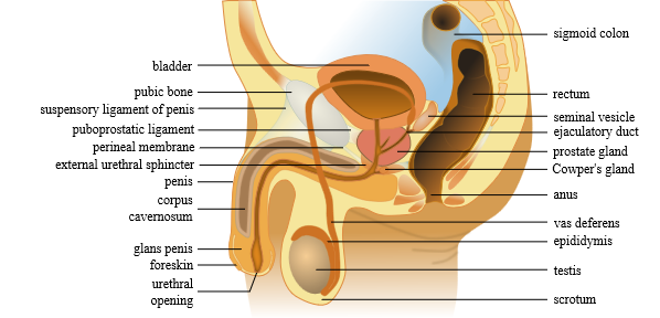
After initial formation in the testes, sperm pass through the following structures, in order:
The epididymis is a whitish mass of tightly coiled tubes against the testicles, acts as a maturation and storage for sperm before they pass into the vas deferens. The vas deferens is a thin tube approximately 12” long that carries the sperm from the epididymis to ejaculatory duct. From the ejaculatory duct, the sperm move through the urethra until their exit from the body. Sperm that do not leave via ejaculation are reabsorbed by the body.
Three accessory glands provide fluids that lubricate the duct system and nourish the sperm cells: seminal vesicles, prostate gland, and bulbourethral glands (Cowper’s gland).
*What is the path of sperm in the male from where it forms to where it leaves the body?
(word bank: ejaculatory duct, epididymis, testes, urethra, vas deferens)
_______________→ ______________→ _______________→ _______________→ _____________
Female Anatomy
____ Observe the female pelvis model, find all numbers/letters indicated on the worksheet at the end of this lab activity.
Major reproductive organs of the female are the ovaries, uterus, and vagina. The female reproductive system involves hormonal communication between key organs, particularly the ovaries and uterus, whose basic functions are to produce the egg, and house the developing embryo, respectively.
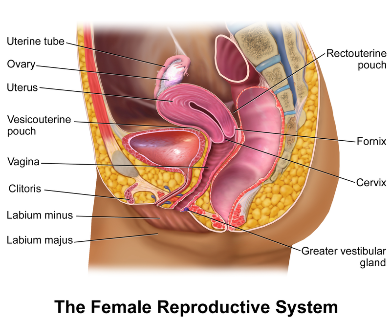
The ovaries are paired and produce both eggs and sex hormones (e.g. estrogen); they are the functional equivalent of the male testes. Ovaries are about the size of a walnut, and at birth contain approximately two million ova (eggs) each. During nearly every menstrual cycle, one (or more) eggs mature and are released during ovulation from the follicle within the ovary, which also releases estrogen. Upon release, the egg passes into a narrow tube known as the oviduct (uterine tube) which is lined with cilia that sweep the egg towards the uterus.
The uterus is fist-sized and thick smooth muscle that contracts during labor & childbirth. The interior lining of the uterus is the endometrium, which thickens during the menstrual cycle in anticipation of housing the developing offspring. The lowermost portion of the uterus narrows into an opening known as the cervix.
The vagina is a muscular organ that serves as a passageway into and out of the uterus. Externally, the female reproductive anatomy (known as the vulva) includes the labia major, labia minor, and clitoris.
Fertilization and Implantation
____ Observe the fertilization model, find the locations of fertilization & implantation, and trace the path of egg and sperm in the female.
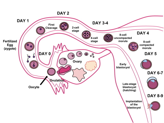
Fertilization & Implantation occur in separate locations and at different times; implantation normally occurs about 8 days post-fertilization. Fertilization (conception) is the process by which the nuclei of a single-celled egg and sperm fuse to form a zygote. Fertilization normally occurs in the oviduct (uterine tube), where the egg travels from the ovary. To reach the egg, sperm cells must travel a distance approximately 100,000 times their body length, which is equivalent to a person running a 110 mile race! Predictably, not all sperm reach the egg, and the first ones that do, generally do not fertilize the egg, since they must penetrate the outer covering of the egg; a single sperm that follows ultimately fuses with the nuclei of the egg to form the zygote.
The zygote is swept by cilia that line the oviduct toward the uterus. As it slowly moves, it undergoes successive rounds of mitosis that build the single cell into a hollow ball of ~100 cells called a blastocyst. After approximately one week, it will reach the uterus where implantation occurs in the blood vessel-rich tissue of the uterus, the endometrium. On occasion, the blastocyst will not reach the uterus and may start to implant on the oviduct. If so, the pregnancy is inviable as this location is not large enough to allow for embryo growth – this is known as an ectopic or tubal pregnancy. An egg, even if fertilized, may also fail to implant in the uterus and then leaves the body during menstruation.
Locations of fertilization and implantation
*Where does fertilization normally occur? ________________________________________
*Where does implantation normally occur? ________________________________________
*What is the path of the egg from where it forms to where it leaves the body if not fertilized?
(word bank: cervix, ovary, oviduct, uterus, vagina)
_______________→ ______________→ _______________→ _______________→ _____________
*What is the path of the sperm from where it enters the female to where it fertilizes the egg?
(word bank: cervix, oviduct, uterus, vagina)
_________________→ ________________→ ________________→ ________________
Early Stages of Development (Chick Embryo)
If the blastocyst successfully implants into the endometrium and continues with mitosis and development, a predictable series of structures form. Three embryonic tissue (or germ) layers (endoderm, mesoderm, ectoderm) are established, and the organ level of development continues until all organs have formed. The first organs to develop are the 1) heart, 2) brain & neural tube, and 3) digestive tract.
____ Examine the early stages of development in the chick embryos under the microscopes. On your worksheet, sketch the stage(s) of the chick embryo and identify/label at least 2 structures.
24 Hour Chick Embryo
Early structures that are visibly forming include somites (blocks of developing muscle tissue), head fold, notochord (below surface), neural groove and fold, and surface ectoderm. See the figure next to the microscope for complete details.
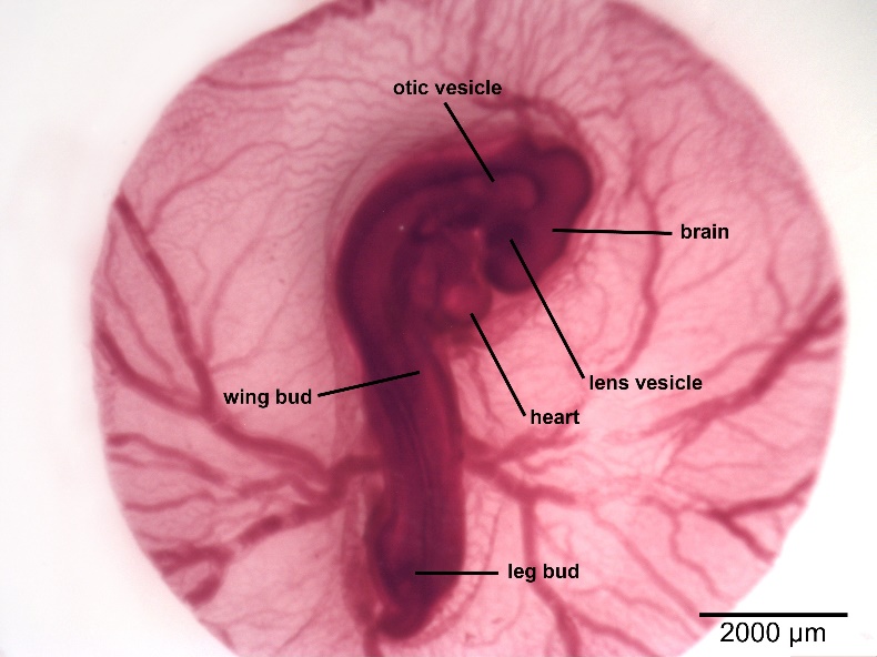
38-43 Hour Chick Embryo
Many organs are now more clearly visible: the heart is now contracting and circulating blood; the brain has several distinct regions (forebrain, midbrain, hindbrain); the eye has a developing lens; the somites enlarge; the neural tube is clearly visible.
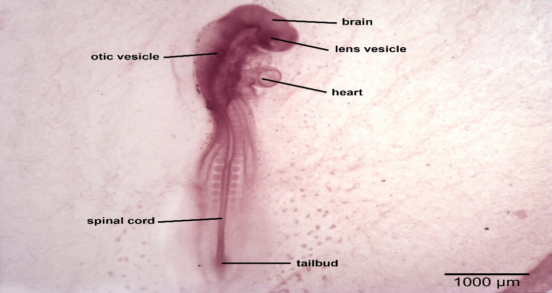
72-Hour Chick Embryo
In addition to the clearly visible heart, eye, brain, and other major organs, a wingbud and hindlimb bud have noticeably formed. The brain has continued to differentiate, while the neural tube has closed along the length of the body—it is now called the spinal cord.
96-Hour Chick Embryo
Tissue differentiation has continued to produce visibly distinct organs. The amnion noticeably surrounds the embryo and the number of somites has increased.
Human Embryonic & Fetal Development
____ Observe & measure the embryo/fetal models on the back table (from 4 wks to 6 mo. gestation).
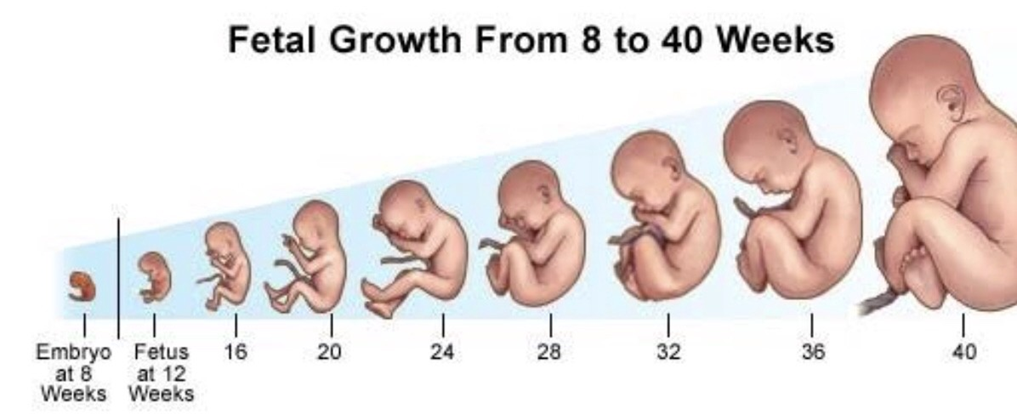
Normal human development requires a full nine months (40 weeks) of gestation to support a fully functioning offspring. Embryo development includes the first eight weeks (two months). By the end of development, all organs have initially formed, but are not functional enough to support independent life. During fetal development (seven months), organs mature and become functional; dramatic weight gain also occurs. The offspring increases greatly from <1” at 4 weeks to about 20” at 38 weeks. It will double in weight from about 4 to 8 pounds between months 7 and 9.
*At _____weeks the baby is 1.5 inches long (use ruler). Size at 5 months (20 weeks)? ____ inches
The Placenta & Pregnancy
Placenta
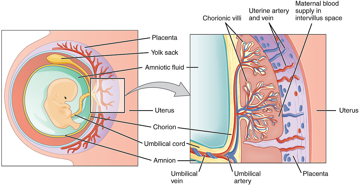
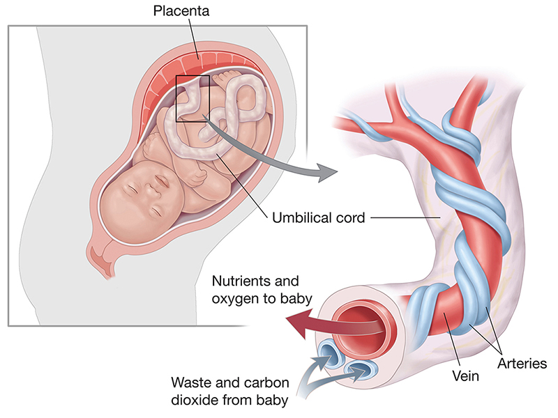
The placenta is a large, blood vessel-rich organ containing tissues from both mom and offspring. It serves as an interface that allows for exchange of nutrients and wastes, while keeping mom and offspring isolated from each other. Blood is not exchanged between mom—offspring, as their bodies may contain incompatible differences (including blood type) between their immune systems.
The fetus is housed in the uterus (womb) and connected to the mother via the umbilical cord which leads to the placenta. The maternal portion of the placenta is the endometrium. The fetal portion of the placenta is the chorion. In addition, the amnion is the layer in which the fetus is housed and surrounded by protective amniotic fluid.
*What materials are exchanged between mother and fetus at the placenta? ________________________
* At the placenta, blood IS / IS NOT exchanged between mother and fetus?
Pregnancy
____ Examine the pregnancy model, be sure to find and name the following structures: 31, 37, 38:
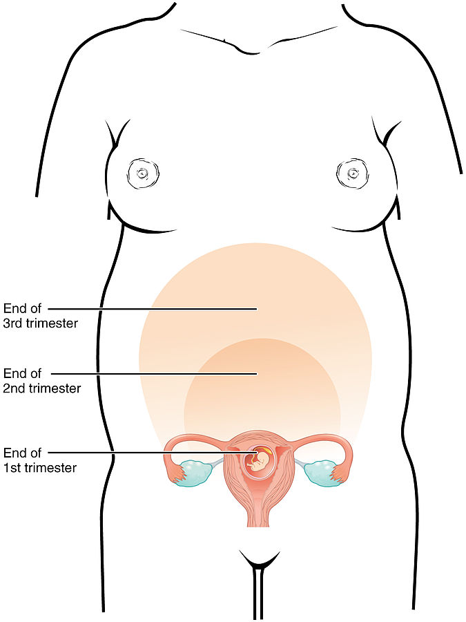
Pregnancy, also known as gestation, lasts approximately 267 days from conception to birth (280 days if calculated from the last menstrual period to childbirth). Pregnancy can be divided into trimesters, each is approximately three months long. The first trimester has the highest possibility of miscarriage (natural death of embryo or fetus). During the second trimester, fetal movements may be felt. At 28 weeks, more than 90% of babies can survive outside of the uterus if provided with high-quality medical care. The third trimester is marked by rapid weight gain.
Major anatomical, metabolic, and physiological changes occur in the mother, including considerable weight gain occurs during a normal pregnancy. As pregnancy progresses, the uterus pushed into the abdominal cavity, exerting pressure on many organs, collecting pushing up against the diaphragm. Physiological changes may include morning sickness, which is thought to be related to elevated hormone levels.
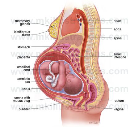
- * Using the pregnancy model, which maternal organs are most compacted by the fetus? _____________
- * Name the two structures (#36-37) the fetus must pass through during birthing: ________, __________
Fetal Circulation Model
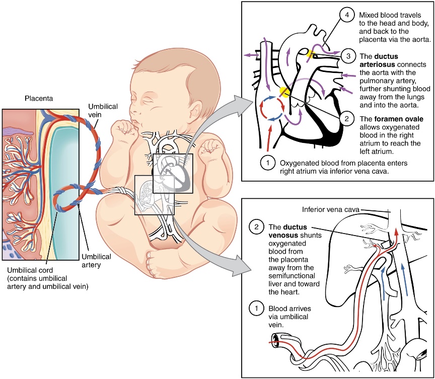
___ Observe the fetal circulatory system model and find the oval opening and the arterial duct.
Two structures in the fetal heart are present that allow blood to bypass a non-utilized organ in the baby—the lungs. In the heart, the oval opening (foramen ovale) between the right atrium and left atrium shunts blood away from the lungs. Also, the arterial duct (ductus arteriosus) connects two blood vessels, the pulmonary trunk and the aorta, to again shunt blood away from the lungs. These structures close after birth in normal development: the oval opening becomes the fossa ovalis, while the arterial duct becomes the ligmentum arteriosum.
* Which two chambers of the heart are connected by the oval opening? ____________, ____________
* The arterial duct connects which two blood vessels? _________________, ____________________
* Which structure is non-functional and being bypassed by the blood? ___________________________
Umbilical Cord
____ Find the umbilical arteries and vein in the model.
The umbilical cord contains a total of 3 blood vessels within it, these facilitate movement of nutrients and wastes between the fetus and the placenta. There are two umbilical arteries going from the fetus to the placenta and one umbilical vein going from the placenta to the fetus (specifically to the liver of the fetus). The umbilical arteries carry wastes away from the fetus including CO2 (they are O2-poor); the umbilical vein brings O2 and nutrients to the fetus. Following birth, the umbilical arteries and vein become non-functional: the umbilical vein becomes the round ligament of the liver, the umbilical arteries regress and become the medial umbilical ligament, and a branch of the anterior division of the internal iliac artery. After it is cut, the remnants of the umbilical cord become the navel or “belly button.”
* Do the umbilical arteries carry O2 rich or O2 poor blood? ____________________________________
* What does the umbilical vein become once the fetus is born? _________________________________
________________________________________________________________________________
Male anatomy By Male_anatomy.png: alt.sex FAQderivative work: Tsaitgaist (talk) – Male_anatomy.png, CC BY-SA 3.0, https://commons.wikimedia.org/w/index.php?curid=6569849By Ttrue12 – Own work, CC BY-SA 3.0, https://commons.wikimedia.org/w/index.php?curid=19679961
Female anatomy By BruceBlaus. When using this image in external sources it can be cited as:Blausen.com staff (2014). “Medical gallery of Blausen Medical 2014”. WikiJournal of Medicine 1 (2). DOI:10.15347/wjm/2014.010. ISSN 2002-4436. – Own work, CC BY 3.0, https://commons.wikimedia.org/w/index.php?curid=29600451
48 hour chick embryo https://sites.newpaltz.edu/histology/developmental-biology/chick/chick-48hr-1x/
72 hour chick embryo https://sites.newpaltz.edu/histology/developmental-biology/chick/chick-72hr-0-6x/
Blastocyst By Seans Potato Business (derivative of the source cited above) – Blastocyst.png, CC BY-SA 3.0, https://commons.wikimedia.org/w/index.php?curid=3306843
Fetal Growth by https://medium.com/@sonal9896225664/stages-of-pregnancy-649b2588423b
Embryo attached to placenta in amniotic cavity By OpenStax College – Anatomy & Physiology, Connexions Web site. http://cnx.org/content/col11496/1.6/, Jun 19, 2013., CC BY 3.0, https://commons.wikimedia.org/w/index.php?curid=30148598
Pregnancy Growth By OpenStax College – Anatomy & Physiology, Connexions Web site. http://cnx.org/content/col11496/1.6/, Jun 19, 2013., CC BY 3.0, https://commons.wikimedia.org/w/index.php?curid=30148608
Pregnancy anatomy by http://baldaivirtuves.info/wp-content/uploads/2017/12/human-anatomy-pregnancy-human-anatomy-pregnancy-anatomy-detail-birth-example-human-free-ideas.jpg
Umbilical cord By http://www.jdimesmedivisual.com/wp-content/gallery/anatomy/BC_Umbilical-Cord_FINAL.jpg
BI 102 Lab Worksheet: Reproductive Name ___________________________ Section _______
1-2. Number the following structures of the reproductive system on the male & female models on your lab table
Male
______ Epididymis
______ Vas deferens
______ Testis
______ Prostate gland
______ Scrotum
______ Urethra
______ Ureter (look at urinary system)
______ Urinary bladder
______ Penis
Female
______ Vagina
______ Oviduct
______ Ovary
______ Urethra
______ Cervix
______ Uterus
______ Urinary bladder
______ Clitoris
______ Rectum
3. In the human female, where does fertilization occur? (where sperm & egg fuse; see model) ______________________
4. In humans, the embryo/fetus develops in which part of the mother’s body (hint: also called the womb)? __________________________
5. Using the pregnancy model at your table, which maternal organs are most compacted by the fetus?
6. Go to the Microscopes: sketch, label & list 2 structures visible in the chick embryo. List embryo stage.
Sketch Structure Name Stage (days or hours)
A.
B.
Go to the back counter:
7. At _____weeks the baby is 1.5 inches long (use ruler). Size at 5 months (20 weeks)? ______ inches
8. In humans, embryonic development occurs during the first ________ months, fetal development occurs during the last ________months.
Go to the front cart:
9. The placenta has a fetal (baby) & maternal (mom) portion. Which forms the fetal portion of the placenta? _______________________________.
Which forms the maternal portion? ______________________________
10. Oval opening (foramen ovale): an opening between which two chambers of the heart? (see model)
_________________________ ________________________________
11. Arterial duct (ductus arteriosis) a blood vessel connecting which two blood vessels?
_______________________________ _____________________________
12. What organ is bypassed by much of the blood in fetal circulation using structures in Q# 10-11 (not used, mom does this task)? ______________________
